

doi: 10.1107/S1744309106027710.
Epub 2006 Jul 26.
Cross-crystallization method used for the crystallization and preliminary diffraction analysis of a novel di-haem cytochrome c4
Affiliations
- PMID: 16880567
- PMCID: PMC2242932
- DOI: 10.1107/S1744309106027710
Item in Clipboard
Cross-crystallization method used for the crystallization and preliminary diffraction analysis of a novel di-haem cytochrome c4
Acta Crystallogr Sect F Struct Biol Cryst Commun.
.
Abstract
The newly discovered di-haem cytochrome c4 from the purple sulfur photosynthetic bacterium Thiocapsa roseopersicina is the first cytochrome c4 to be crystallized from an anaerobic organism. It was crystallized using the addition of metal-ion salts to the standard vapour-diffusion method. Coloured well shaped three-dimensional crystals with dimensions of approximately 0.6 x 0.05 x 0.02 mm grew within 3-4 d at pH 5 and diffracted to 1.72 angstroms without radiation damage. Cytochrome c4 crystallized in space group P4(1)2(1)2 as a primitive tetragonal system with unit-cell parameters a = b = 75.29, c = 37.12 angstroms, alpha = beta = gamma = 90 degrees.
Figures
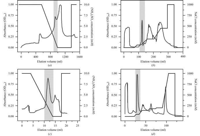
Purification of cytochrome c
4 from the purple photosynthetic bacteria T. roseopersicina. (a) Butyl-Sepharose chromatography. (b) Q-Sepharose chromatography. (c) Phenyl-Sepharose chromatography. (d) Q-Sepharose chromatography. Shaded areas represent cytochrome-containing fractions. For experimental details see text.

Pseudocrystals of cytochrome c
4 grown from solution containing 1.7 M (NH4)2SO4, 100 mM sodium citrate pH 6.0 and 100 mM NaCl.
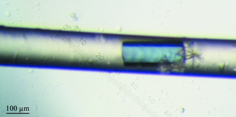
Crystal of cytochrome c
4 grown inside the 0.1 mm capillary using the advanced crystallization method based on counter-diffusion.
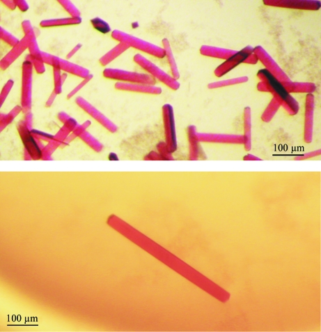
Colored well shaped three-dimensional crystals of cytochrome c
4 with maximal dimensions of approximately 0.6 × 0.05 × 0.02 mm grown within 3–4 d in the presence of 5 mM CuCl2, 3.2 M (NH4)2SO4 and 0.1 M citric acid pH 5 in the protein drop and 3.2 M (NH4)2SO4, 0.1 M citric acid pH 5 in the reservoir and with the influence of the additives 5 mM CdCl2, 5 mM CoCl2, 5 mM BaCl2 in the other wells (see also §3.1).
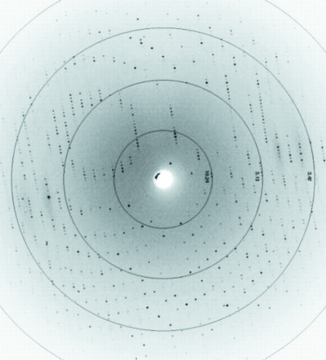
Diffraction image of a cytochrome c
4 crystal collected at the EMBL Hamburg Outstation at beamline X11 to a resolution of 1.72 Å.
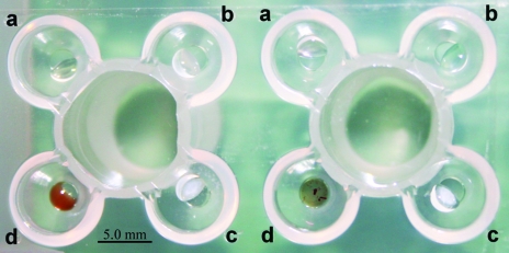
Emerald Biosystems crystallization plate (JenaBioscience, Germany) used to perform the cross-crystallization procedure. Drop wells a, b, c and d were each filled with 0.5 µl of various 5 mM chloride salts (see text) and 0.5 µl precipitant. Protein at a concentration of 15 mg ml−1 (1 µl) was only added into drop well d, containing 5 mM CuCl2.
References
-
- Asherie, N. (2004). Methods, 34, 266–272. - PubMed
-
- Bagyinka, C., Ösz, J. & Szaraz, S. (2003). J. Biol. Chem.278, 20624–20627. - PubMed
-
- Benini, S., Gonzáles, A., Rypniewski, W. R., Wilson, K. S., Van Beeumen, J. J. & Ciurli, S. (2000). Biochemistry, 39, 13115–13126. - PubMed
-
- Bergfors, T. M. (1999). Protein Crystallization: Techniques, Strategies and Tips. La Jolla, CA, USA: International University Line.
Publication types
MeSH terms
Substances
LinkOut - more resources
Full Text Sources
Miscellaneous
