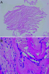
Cholesteatoma of the clivus
- PMID: 16880901
- PMCID: PMC1408076
- DOI: 10.1055/s-2006-926218
Cholesteatoma of the clivus
Abstract
Cholesteatomas (central nervous system epidermoids) can be found intradurally or extradurally in the central nervous system. Extradural intraosseous lesions are most commonly found in the petrous bone. The authors describe a unique case of a clival cholesteatoma in a 64-year-old woman who presented with headaches. No other neurological complaints or physical examination findings were noted. Magnetic resonance imaging showed an expansile lesion centered in the middle portion of the clivus. A large portion of the clivus was eroded. The lesion was explored via a transnasal trans-sphenoidal approach and granular debris was evacuated. The cystic lining was stripped from the surrounding bone, and the bone opening was widely fenestrated. Pathological examination showed keratinous debris with macrophages and an outer lining of benign epithelial tissue consistent with a cholesteatoma (epidermoid cyst). When surgically accessible, these lesions should be excised to prevent a recurrence. If inaccessible, marsupialization may be considered.
Figures


Similar articles
-
[Endoscopic transnasal approach in surgical treatment of petrous temporal bone cholesteatoma extending towards the clivus. Three clinical observations and literature review].Zh Vopr Neirokhir Im N N Burdenko. 2022;86(2):97-102. doi: 10.17116/neiro20228602197. Zh Vopr Neirokhir Im N N Burdenko. 2022. PMID: 35412718 Review. Russian.
-
[Two cases of the epidermoid on the petrous bone].No Shinkei Geka. 2000 Sep;28(9):797-802. No Shinkei Geka. 2000. PMID: 11025879 Japanese.
-
Retraction pockets and attic cholesteatomas.Acta Otorhinolaryngol Belg. 1980;34(1):62-84. Acta Otorhinolaryngol Belg. 1980. PMID: 6162358
-
Chondroblastoma of the Clivus: Case Report and Review.J Neurol Surg Rep. 2015 Nov;76(2):e258-64. doi: 10.1055/s-0035-1564601. Epub 2015 Oct 9. J Neurol Surg Rep. 2015. PMID: 26623238 Free PMC article.
-
Intracranial cholesteatoma - case report and critical review.Clin Neuropathol. 2009 Nov-Dec;28(6):440-4. doi: 10.5414/npp28440. Clin Neuropathol. 2009. PMID: 19919818 Review.
Cited by
-
Endoscopic Transnasal and Transclival Resection of Cholesteatomas in the Clivus and Temporal Bone Pyramid: A Case Series of Eight Patients.Cureus. 2025 Jan 12;17(1):e77330. doi: 10.7759/cureus.77330. eCollection 2025 Jan. Cureus. 2025. PMID: 39935915 Free PMC article.
-
Surgical Guidance for Removal of Cholesteatoma Using a Multispectral 3D-Endoscope.Sensors (Basel). 2020 Sep 17;20(18):5334. doi: 10.3390/s20185334. Sensors (Basel). 2020. PMID: 32957675 Free PMC article.
-
Determination of the optical properties of cholesteatoma in the spectral range of 250 to 800 nm.Biomed Opt Express. 2020 Feb 20;11(3):1489-1500. doi: 10.1364/BOE.384742. eCollection 2020 Mar 1. Biomed Opt Express. 2020. PMID: 32206424 Free PMC article.
References
-
- Axon P R, Fergie N, Saeed S R, Temple R H, Ramsden R T. Petrosal cholesteatoma: management considerations for minimizing morbidity. Am J Otol. 1999;20:505–510. - PubMed
-
- Horn K L. Intracranial extension of acquired aural cholesteatoma. Laryngoscope. 2000;110:761–772. - PubMed
-
- Pisaneschi M J, Langer B. Congenital cholesteatoma and cholesterol granuloma of the temporal bone: role of magnetic resonance imaging. Top Magn Reson Imaging. 2000;11:87–97. - PubMed

