

Inductive tissue engineering with protein and DNA-releasing scaffolds
- PMID: 16880921
- PMCID: PMC2657198
- DOI: 10.1039/b514174p
Inductive tissue engineering with protein and DNA-releasing scaffolds
Abstract
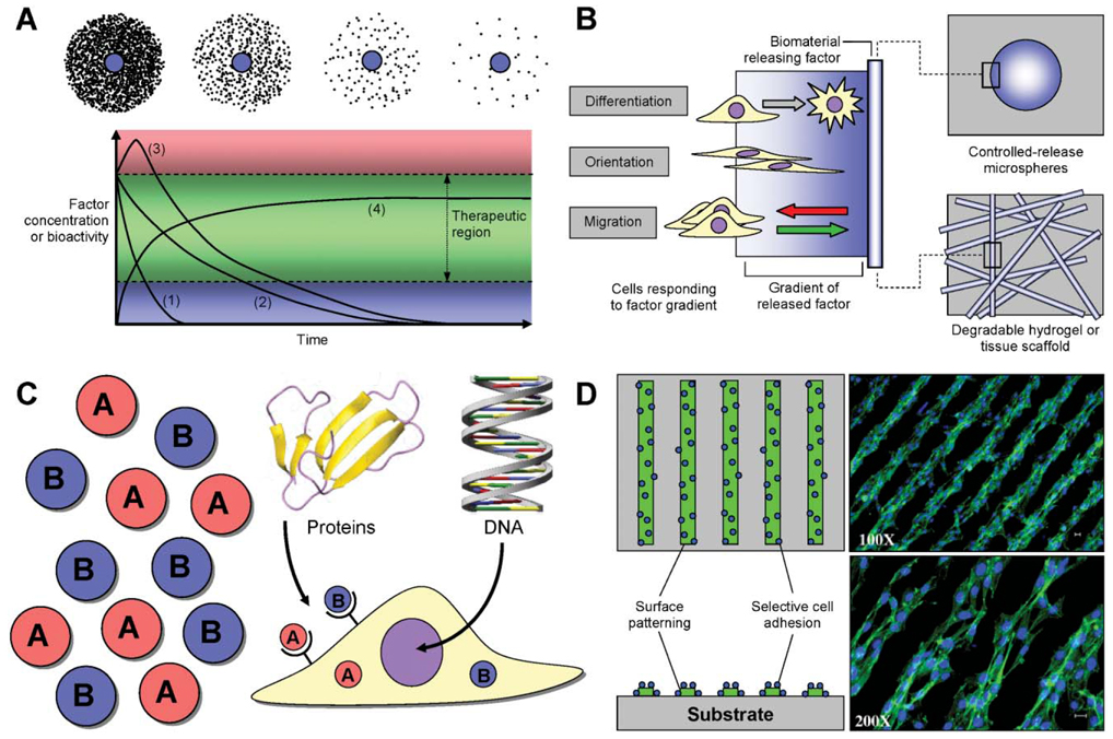
Cellular differentiation, organization, proliferation and apoptosis are determined by a combination of an intrinsic genetic program, matrix/substrate interactions, and extracellular cues received from the local microenvironment. These molecular cues come in the form of soluble (e.g. cytokines) and insoluble (e.g. ECM proteins) factors, as well as signals from surrounding cells that can promote specific cellular processes leading to tissue formation or regeneration. Recent developments in the field of tissue engineering have employed biomaterials to present these cues, providing powerful tools to investigate the cellular processes involved in tissue development, or to devise therapeutic strategies based on cell replacement or tissue regeneration. These inductive scaffolds utilize natural and/or synthetic biomaterials fabricated into three-dimensional structures. This review summarizes the use of scaffolds in the dual role of structural support for cell growth and vehicle for controlled release of tissue inductive factors, or DNA encoding for these factors. The confluence of molecular and cell biology, materials science and engineering provides the tools to create controllable microenvironments that mimic natural developmental processes and direct tissue formation for experimental and therapeutic applications.
Figures

Similar articles
-
Three-Dimensional Biomaterials with Spatiotemporal Control for Regenerative Tissue Engineering.Acc Chem Res. 2023 Jun 6;56(11):1313-1319. doi: 10.1021/acs.accounts.2c00666. Epub 2023 Apr 27. Acc Chem Res. 2023. PMID: 37103937
-
Nanotopographical Features of Polymeric Nanocomposite Scaffolds for Tissue Engineering and Regenerative Medicine: A Review.Biomimetics (Basel). 2025 May 15;10(5):317. doi: 10.3390/biomimetics10050317. Biomimetics (Basel). 2025. PMID: 40422147 Free PMC article. Review.
-
Nanocomposite injectable gels capable of self-replenishing regenerative extracellular microenvironments for in vivo tissue engineering.Biomater Sci. 2018 Feb 27;6(3):550-561. doi: 10.1039/c7bm01167a. Biomater Sci. 2018. PMID: 29379910
-
Engineering hydrogels as extracellular matrix mimics.Nanomedicine (Lond). 2010 Apr;5(3):469-84. doi: 10.2217/nnm.10.12. Nanomedicine (Lond). 2010. PMID: 20394538 Free PMC article. Review.
-
Natural-Based Hydrogels for Tissue Engineering Applications.Molecules. 2020 Dec 11;25(24):5858. doi: 10.3390/molecules25245858. Molecules. 2020. PMID: 33322369 Free PMC article. Review.
Cited by
-
Emerging technologies for assembly of microscale hydrogels.Adv Healthc Mater. 2012 Mar;1(2):149-158. doi: 10.1002/adhm.201200011. Adv Healthc Mater. 2012. PMID: 23184717 Free PMC article. Review.
-
Constraining the Pluripotent Fate of Human Embryonic Stem Cells for Tissue Engineering and Cell Therapy - The Turning Point of Cell-Based Regenerative Medicine.Br Biotechnol J. 2013 Oct 1;3(4):424-457. doi: 10.9734/BBJ/2013/4309#sthash.6D8Rulbv.dpuf. Br Biotechnol J. 2013. PMID: 24926434 Free PMC article.
-
Biomolecule gradient in micropatterned nanofibrous scaffold for spatiotemporal release.Langmuir. 2012 Sep 25;28(38):13675-87. doi: 10.1021/la302386u. Epub 2012 Sep 14. Langmuir. 2012. PMID: 22950580 Free PMC article.
-
Bridging the lesion-engineering a permissive substrate for nerve regeneration.Regen Biomater. 2015 Sep;2(3):203-14. doi: 10.1093/rb/rbv012. Epub 2015 Aug 10. Regen Biomater. 2015. PMID: 26816642 Free PMC article.
-
Matrices and scaffolds for DNA delivery in tissue engineering.Adv Drug Deliv Rev. 2007 May 30;59(4-5):292-307. doi: 10.1016/j.addr.2007.03.017. Epub 2007 Apr 14. Adv Drug Deliv Rev. 2007. PMID: 17512630 Free PMC article. Review.
References
-
- Langer R, Vacanti JP. Tissue engineering. Science. 1993;260(5110):920–926. - PubMed
-
- Wobus AM, Boheler KR. Embryonic stem cells: prospects for developmental biology and cell therapy. Physiol. Rev. 2005;85(2):635–678. - PubMed
-
- Keller G. Embryonic stem cell differentiation: emergence of a new era in biology and medicine. Genes Dev. 2005;19(10):1129–1155. - PubMed
-
- Murphy WL, Mooney DJ. Controlled delivery of inductive proteins, plasmid DNA and cells from tissue engineering matrices. J. Periodontal Res. 1999;34(7):413–419. - PubMed
Publication types
MeSH terms
Substances
Grants and funding
LinkOut - more resources
Full Text Sources
Other Literature Sources