. 2006;8(4):R49.
doi: 10.1186/bcr1538.

Estrogen receptor-alpha and progesterone receptor are expressed in label-retaining mammary epithelial cells that divide asymmetrically and retain their template DNA strands

Affiliations

Estrogen receptor-alpha and progesterone receptor are expressed in label-retaining mammary epithelial cells that divide asymmetrically and retain their template DNA strands

Brian W Booth et al. Breast Cancer Res. 2006.

Abstract

Introduction: Stem cells of somatic tissues are hypothesized to protect themselves from mutation and cancer risk through a process of selective segregation of their template DNA strands during asymmetric division. Mouse mammary epithelium contains label-retaining epithelial cells that divide asymmetrically and retain their template DNA.

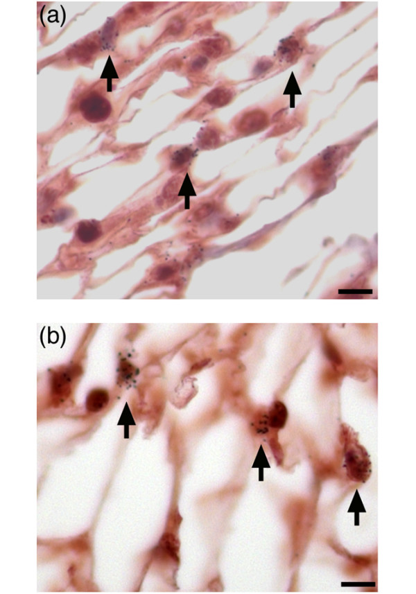
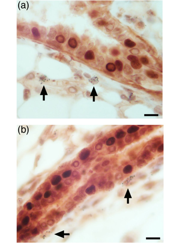
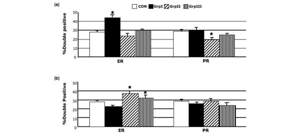
Method: Immunohistochemistry was used in murine mammary glands that had been labeled with [3H]thymidine during allometric growth to investigate the co-expression of DNA label retention and estrogen receptor (ER)-alpha or progesterone receptor (PR). Using the same methods, we investigated the co-localization of [3H]thymidine and ER-alpha or PR in mammary tissue from mice that had received treatment with estrogen, progesterone, and prolactin subsequent to a long chase period to identify label-retaining cells.

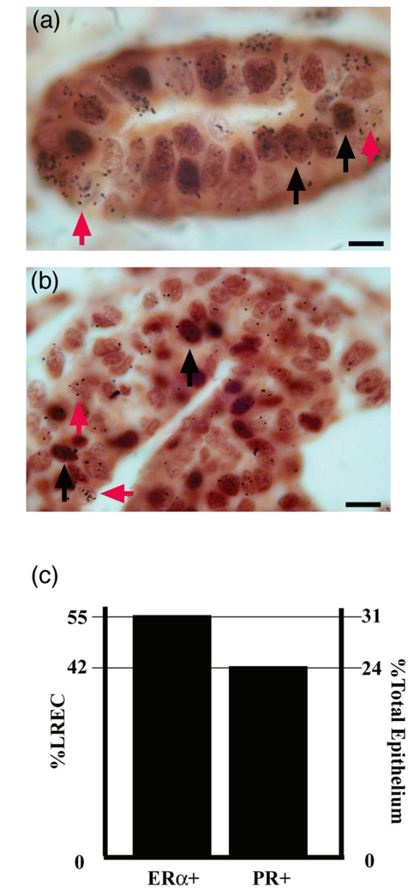
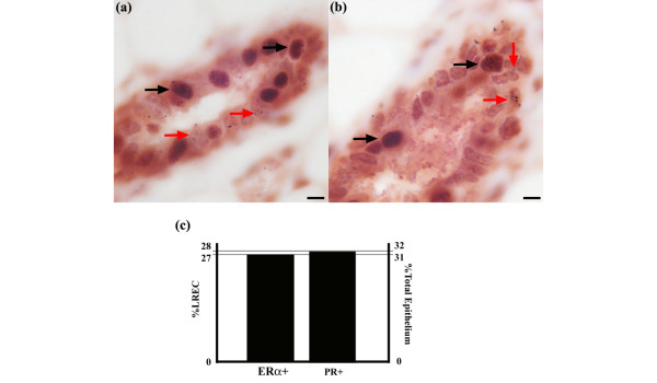
Results: Label-retaining epithelial cells (LRECs) comprised approximately 2.0% of the entire mammary epithelium. ER-alpha-positive and PR-positive cells represented about 30-40% of the LREC subpopulation. Administration of estrogen, progesterone, and prolactin altered the percentage of LRECs expressing ER-alpha.

Conclusion: The results presented here support the premise that there is a subpopulation of LRECs in the murine mammary gland that is positive for ER-alpha and/or PR. This suggests that certain mammary LRECs (potentially stem cells) remain stably positive for these receptors, raising the possibility that LRECs comprise a hierarchy of asymmetrically cycling mammary stem/progenitor cells that are distinguished by the presence or absence of nuclear steroid receptor expression.


Figures

Figure 1
Figure 1
ER-α-positive and PR-positive epithelial cells are in cycle in during allometric growth. (a) ER-α and [3H]thymidine are detected in mammary tissue. ER-α-positive staining is indicated with black arrowheads and [3H]thymidine incorporation is indicated with red arrowheads. (b) PR and [3H]thymidine are detected in mammary tissue. PR-positive staining is indicated with black arrowheads and [3H]thymidine incorporation is indicated with red arrowheads. (c) Graphical depiction of the number of cells that were double positive for [3H]thymidine incorporation and ER-α or PR (left scale) and the percentage of cells that were ER-α or PR positive within epithelium. Scale bars = 5 μm. ER, estrogen receptor; PR, progesterone receptor.
Figure 2
Figure 2
Stromal cells proliferate in pubertal mammary tissue. The nuclear label [3H]thymidine is detected in mammary tissue that has been processed for (a) ER-α or (b) PR. Black arrows designate stromal cells that are [3H]thymidine positive, indicating cells that have recently undergone cell cycle. Scale bars = 5 μm. ER, estrogen receptor; PR, progesterone receptor.
Figure 3
Figure 3
A subpopulation of LRECs is ER-α positive or PR positive. (a) ER-α and [3H]thymidine are detected in mammary tissue. LRECs that are double positive for ER-α and [3H]thymidine incorporation are indicated with black arrows. Cells that are LRECs alone are indicated with red arrowheads. (b) PR and [3H]thymidine are detected in mammary tissue. LRECs that are double positive for PR and [3H]thymidine incorporation are indicated with black arrows. Cells that are LRECs alone are indicated with red arrows. (c) Graphical depiction of the number of cells that are double positive for [3H]thymidine incorporation and ER-α or PR (left scale) and percentage of cells that are ER-α or PR positive within epithelium. Scale bars = 5 μm. ER, estrogen receptor; LREC, label-retaining epithelial cell; PR, progesterone receptor.
Figure 4
Figure 4
Periductal stromal cells retain nuclear label. The nuclear label [3H]thymidine is detected in mammary tissue that has been processed for (a) ER-α or (b) PR following a 7-week chase period. Black arrows indicate periductal stromal cells that are [3H]thymidine positive. Scale bars = 5 μm. ER, estrogen receptor; PR, progesterone receptor.
Figure 5
Figure 5
Graphical depiction of percentage of LRECs that were either ER-α positive or PR positive after hormone treatments. All animals received [3H]thymidine at 5 weeks of age. Control animals (Con; white bar) received only [3H]thymidine. After a 7-week chase group I (GrpI; black bar) received estradiol injections; group II (GrpII; diagonal hatches) animals received estradiol and progesterone; and group III (GrpIII; vertical hatches) received estradiol, progesterone, and prolactin. (a) Results after 3-day hormone treatment and (b) results obtained after 5-day hormone treatment and 2-day recovery period. Error bars indicate the standard error of the mean. *P < 0.05, versus Con. ER, estrogen receptor; LREC, label-retaining epithelial cell; PR, progesterone receptor.

References

    1. Cairns J. Mutation selection and the natural history of cancer. Nature. 1975;255:197–200. doi: 10.1038/255197a0. - DOI - PubMed
    1. Potten CS, Hume WJ, Reid P, Cairns J. The segregation of DNA in epithelial stem cells. Cell. 1978;15:899–906. doi: 10.1016/0092-8674(78)90274-X. - DOI - PubMed
    1. Potten CS, Owen G, Booth D. Intestinal stem cells protect their genome by selective segregation of template DNA strands. J Cell Sci. 2002;115:2381–2388. - PubMed
    1. Smith GH. Label-retaining epithelial cells in mouse mammary gland divide asymmetrically and retain their template DNA strands. Development. 2005;132:681–687. doi: 10.1242/dev.01609. - DOI - PubMed
    1. Anderson E, Clarke RB. Steroid receptors and cell cycle in normal mammary epithelium. J Mammary Gland Biol Neoplasia. 2004;9:3–13. doi: 10.1023/B:JOMG.0000023584.01750.16. - DOI - PubMed

Publication types