. 2006 Aug 8;103(32):12057-62.
doi: 10.1073/pnas.0604247103. Epub 2006 Aug 1.

Reconstitution of paired T cell receptor alpha- and beta-chains from microdissected single cells of human inflammatory tissues

Affiliations

Reconstitution of paired T cell receptor alpha- and beta-chains from microdissected single cells of human inflammatory tissues

Sabine Seitz et al. Proc Natl Acad Sci U S A. .

Abstract

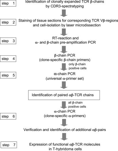
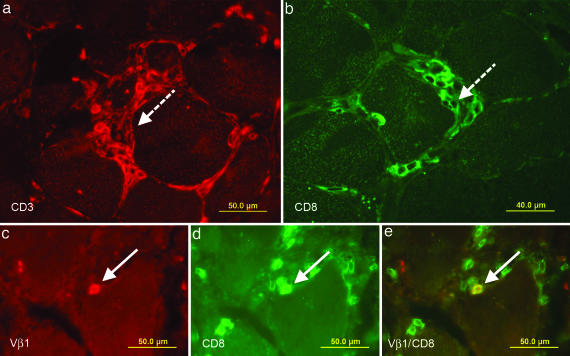
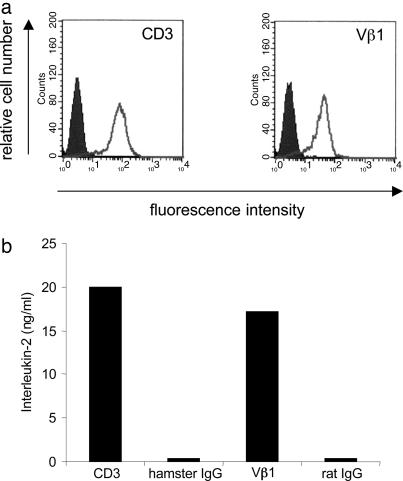
We describe a strategy to "revive" putatively pathogenic T cells from frozen specimens of human inflammatory target organs. To distinguish pathogenic from irrelevant bystander T cells, we focused on cells that were (i) clonally expanded and (ii) in direct morphological contact with a target cell. Using CDR3 spectratyping, we identified clonally expanded T cell receptor (TCR) beta-chains in muscle sections of patients with inflammatory muscle diseases. By immunohistochemistry, we identified those Vbeta-positive T cells that fulfilled the morphological criteria of myocytotoxicity and isolated them by laser microdissection. Next, we identified coexpressed pairs of TCR alpha- and beta-chains by a multiplex PCR protocol, which allows the concomitant amplification of both chains from single cells. This concomitant amplification had not been achieved previously in histological sections, mainly because of the paucity of available anti-alpha-chain antibodies and the great heterogeneity of the alpha-chain genes. From muscle tissue of a patient with polymyositis, we isolated 64 T cells that expressed an expanded Vbeta1 chain. In 23 of these cells, we identified the corresponding alpha-chain. Twenty of these 23 alpha-chains were identical, suggesting antigen-driven selection. After functional reconstitution of the alphabeta-pairs, their antigen-recognition properties could be studied. Our results open avenues for combined analysis of the full TCR alpha- and beta-chain repertoire in human inflammatory tissues.


Conflict of interest statement

Conflict of interest statement: No conflicts declared.

Figures

Fig. 1.
Fig. 1.
Strategy to identify paired TCR α- and β-chains from microdissected tissue-infiltrating T cells. In the first step, we investigated clonal T cell expansions by CDR3 spectratyping of the TCR β-chains. Then, in step 2, we stained sections from frozen biopsy specimens with appropriate anti-Vβ antibodies and isolated positive cells by microdissection. After cDNA preparation and a preamplification PCR for α- and β-chains (step 3), we tested the microdissected cells for expression of the expanded β-chain by PCR using clone-specific β-primers (step 4). Cells that expressed the correct sequence, i.e., the sequence identified by CDR3 spectratyping, were examined for the matching α-chains with a universal primer set that allows amplification of all TCR α-chain sequences (step 5). The preamplification product served as template. Based on the α-chain sequences identified in step 5, we designed clone-specific α-chain primers and reexamined all β-chain-positive cells (step 6), which allowed identification of additional αβ-pairs because clone-specific primers are more efficient than the universal primer set. For functional studies, the corresponding α- and β-chains were expressed on the surface of a T hybridoma cell line (step 7).
Fig. 2.
Fig. 2.
Immunolocalization of T cells in muscle tissue of patient PM16488. Cryosections of a frozen biopsy specimen were stained in red with a Cy3-labeled antibody to CD3 (a) and in green with an FITC-labeled antibody to CD8 (b). Dense focal invasions of T cells into muscle fibers are indicated in both figures by dashed arrows. Focal invasions are one of the diagnostic criteria of PM. However, such closely packed T cell aggregates are not suited for the isolation of single T cells by microdissection. We therefore focused on distinct single cells that indent or directly contact a muscle fiber. (ce) Double stainings of TCR Vβ1 and CD8. Biopsy sections were stained in red with a Cy3-labeled anti-Vβ1 antibody (c) and in green with an FITC-labeled anti-CD8 antibody (d). (e) A double-positive cell in direct contact with a muscle fiber appears in yellow and is indicated by an arrow.
Fig. 3.
Fig. 3.
Characterization of the Vα2.2-Vβ1-TCR reconstituted in the T hybridoma cell line 58αβ. (a) Cell-surface expression as determined by flow cytometry. The TCR transfectants were stained positive with antibodies to mouse CD3 (a Left) and human TCR Vβ1 (a Right) molecules (white areas). The transfectants were not stained with the corresponding isotype control antibodies hamster IgG and rat IgG, respectively (shaded areas). Clone A5 is shown as a representative of three independent clones. These data provide evidence that the reconstituted TCR is expressed at the cell surface of 58αβ cells. (b) Antibody-induced activation of the reconstituted Vα2.2-Vβ1-TCR clone A5. Antibodies to mouse CD3, Vβ1, and the corresponding isotype control antibodies were coated on microtiter plates, the TCR transfectants were added, and IL-2 was measured in the supernatants by ELISA. The transfectants produced IL-2 in response to anti-CD3 and anti-Vβ1 antibodies but not to the respective control antibodies. These results show that the reconstructed, revived TCR is functional.

References

    1. Steinman L. J. Exp. Med. 2003;197:1065–1071. - PMC - PubMed
    1. Cohen I. R. Immunol. Today. 1992;13:490–494. - PubMed
    1. Bach J.-F., Chatenoud L. Annu. Rev. Immunol. 2001;19:131–161. - PubMed
    1. von Herrath M. Nature. 2005;435:151–152. - PubMed
    1. Krogsgaard M., Davis M. M. Nat. Immunol. 2005;6:239–245. - PubMed

Publication types

Substances

LinkOut - more resources