. 2006;2006(2):68091.
doi: 10.1155/JBB/2006/68091.

Cloning and expression of human membrane-bound and soluble engineered T cell receptors for immunotherapy

Affiliations

Cloning and expression of human membrane-bound and soluble engineered T cell receptors for immunotherapy

Nehad M Alajez et al. J Biomed Biotechnol. 2006.

Abstract

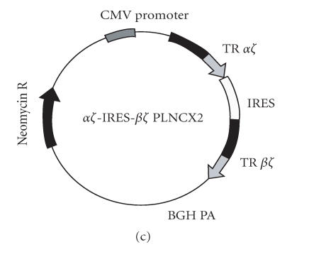
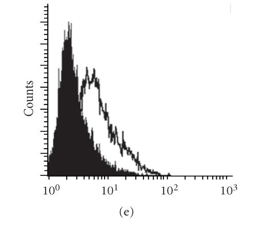
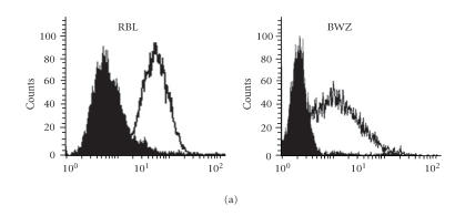
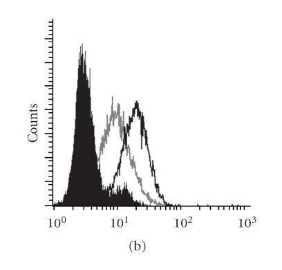
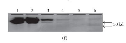
We report here the design and construction of several gene vectors for expression in mammalian cells of membrane-bound and soluble human T cell receptors (TR). We designed a vector (TR-ALPHA-IRES-TR-BETA pEF4) that encodes high-level expression of the full-length TR on the surface of T cells. Furthermore, we engineered TR that does not require the presence of endogenous CD3 molecules for surface expression and thus expression is not limited to T cells. We also constructed a vector encoding a single-chain TR (scTR) as a fusion protein of V-ALPHA-V-BETA-C-BETA with CD3Z. Since it is encoded and expressed as a single molecule, this scTR is well suited for gene therapy. Lastly, we successfully used a mammalian expression vector for generation of soluble human TR. The approaches we used here for manipulation of a human tumor-specific TR can be useful for other investigators interested in TR-based immunotherapy.


Figures

Figure 1
Figure 1
Expression of the TRAZ from MA CTL clone on the surface of a TR-deficient Jurkat line (JRT3-T3.5). (a) The TR-ALPHA-IRES-TR-BETA cassette was cloned into the pEF4 expression vector. (b) Untransfected JRT3-T3.5, (c) JRT3-T3.5 cells transfected with the TCRB sequence, or (d) JRT3-T3.5 cells transfected with MA TR-ALPHA-IRES-TR-BETA pEF4 were stained with anti-CD3 Epsilon (open histogram) or with isotype control (filled histogram). IRES stands for internal ribosomal entry site. Zeocin is antibiotic resistance gene.
Figure 1
Figure 1
Expression of the TRAZ from MA CTL clone on the surface of a TR-deficient Jurkat line (JRT3-T3.5). (a) The TR-ALPHA-IRES-TR-BETA cassette was cloned into the pEF4 expression vector. (b) Untransfected JRT3-T3.5, (c) JRT3-T3.5 cells transfected with the TCRB sequence, or (d) JRT3-T3.5 cells transfected with MA TR-ALPHA-IRES-TR-BETA pEF4 were stained with anti-CD3 Epsilon (open histogram) or with isotype control (filled histogram). IRES stands for internal ribosomal entry site. Zeocin is antibiotic resistance gene.
Figure 2
Figure 2
Construction and expression of engineered MUC1-specific TRAZ and TRBZ receptors. Expression vectors for (a) TRAZ, (b) BZ, and (c) AZ/BZ. (d) Untransfected 293H cells, (e) 293H cells cotransfected with the TRAZ and TRBZ, or (f) cells transfected with the AZ-IRES-BZ pLNCX2 were stained for surface expression of the TR using anti-TR antibody BF1 (open histogram) or isotype control antibody (filled histogram).
Figure 2
Figure 2
Construction and expression of engineered MUC1-specific TRAZ and TRBZ receptors. Expression vectors for (a) TRAZ, (b) BZ, and (c) AZ/BZ. (d) Untransfected 293H cells, (e) 293H cells cotransfected with the TRAZ and TRBZ, or (f) cells transfected with the AZ-IRES-BZ pLNCX2 were stained for surface expression of the TR using anti-TR antibody BF1 (open histogram) or isotype control antibody (filled histogram).
Figure 3
Figure 3
Construction and expression of MUC1-specific single-chain T cell receptors (scTRs). (a) 293H cells were transfected with (b) the scTR, (c) scTR-CD4TM-hZ, or (d) scTR-CD4TM-AGD-hZ mammalian expression vectors. Cells were stained with anti-TR BF1 (open histogram) or isotype control (filled histogram) antibody. (e) shows quantitative comparison of TR expression on 293H cells transfected with different scTR constructs. P < .05.
Figure 3
Figure 3
Construction and expression of MUC1-specific single-chain T cell receptors (scTRs). (a) 293H cells were transfected with (b) the scTR, (c) scTR-CD4TM-hZ, or (d) scTR-CD4TM-AGD-hZ mammalian expression vectors. Cells were stained with anti-TR BF1 (open histogram) or isotype control (filled histogram) antibody. (e) shows quantitative comparison of TR expression on 293H cells transfected with different scTR constructs. P < .05.
Figure 4
Figure 4
Expression of functional scTR on the surface of T and non-T immune cells. (a) Rat basophilic leukemia (RBL) or BWZ cells were transfected with the scTR-pEF6 and were stained for surface expression with anti-TR BF1 antibody (open) or with isotype control (filled) histogram. (b) IL-2 secretion from BWZ cells (open bars) or BWZ-scTR (filled bars) following stimulated with SEE superantigen or with anti-TR BF1 antibody. Stimulation with Ionomycin/PMA (I/P) served as the positive control.
Figure 4
Figure 4
Expression of functional scTR on the surface of T and non-T immune cells. (a) Rat basophilic leukemia (RBL) or BWZ cells were transfected with the scTR-pEF6 and were stained for surface expression with anti-TR BF1 antibody (open) or with isotype control (filled) histogram. (b) IL-2 secretion from BWZ cells (open bars) or BWZ-scTR (filled bars) following stimulated with SEE superantigen or with anti-TR BF1 antibody. Stimulation with Ionomycin/PMA (I/P) served as the positive control.
Figure 5
Figure 5
Expression and purification of soluble scTR following surface biotin labeling and immunoprecipitation. (a) scTR expression vector encoding a thrombin cleavage site, T. (b) RBL cells transfected with the scTR were stained with anti-TR BF1 antibody (open histograms) before (right) or after (left) treatment with thrombin. Filled histogram shows staining with isotype control antibody. (c) Immunoprecipitation of the scTR from RBL (lanes 1 and 3) or RBL cells transfected with the scTR (lanes 2 and 4) before (lanes 1 and 2) or after (lanes 3 and 4) treatment with thrombin. Lanes 6–8 are SA-HRP blotting of fraction eluted with 150 mM Glycine, PH 2.2, 100 mM Glycine PH 2.2, or diethylamine (DEA) PH 11.2, respectively. Lane 5 is IP from control lysate.
Figure 5
Figure 5
Expression and purification of soluble scTR following surface biotin labeling and immunoprecipitation. (a) scTR expression vector encoding a thrombin cleavage site, T. (b) RBL cells transfected with the scTR were stained with anti-TR BF1 antibody (open histograms) before (right) or after (left) treatment with thrombin. Filled histogram shows staining with isotype control antibody. (c) Immunoprecipitation of the scTR from RBL (lanes 1 and 3) or RBL cells transfected with the scTR (lanes 2 and 4) before (lanes 1 and 2) or after (lanes 3 and 4) treatment with thrombin. Lanes 6–8 are SA-HRP blotting of fraction eluted with 150 mM Glycine, PH 2.2, 100 mM Glycine PH 2.2, or diethylamine (DEA) PH 11.2, respectively. Lane 5 is IP from control lysate.
Figure 6
Figure 6
Expression and purification of soluble scTR using mammalian expression system. (a) The single-chain fraction variable (scFv) domain was cloned and fused to a C-terminus HA and c-myc epitope tags. (b) Secreted scTCR that was cloned and fused to a C-terminus Flag and 6-His epitope tags. (c) and (d) the secreted scTR as described in (b), with the exception that the secreted scTR was fused to the leader sequence from GM-CSF (c) or from Ig-κ light chain (d). (e) Western blot of the culture supernatants from 293H cells transiently transfected with constructs a–d (a′–d′), immunoprecipitated with appropriate anti-tag antibody and blotted with anti-c-myc antibody (a′) or with anti-Flag-M2 antibody (b′–d′). Ctr is supernatant from untransfected cells. (f) Coomassie blue staining of fractions from culture supernatant b′ purified using nickel column. Lane 1 is culture supernatant before purification, 2 is flow through, 3 is wash, and 4–6 are different eluted fractions. (g) Western blot of panel (f) using anti-Flag M2 antibody.
Figure 6
Figure 6
Expression and purification of soluble scTR using mammalian expression system. (a) The single-chain fraction variable (scFv) domain was cloned and fused to a C-terminus HA and c-myc epitope tags. (b) Secreted scTCR that was cloned and fused to a C-terminus Flag and 6-His epitope tags. (c) and (d) the secreted scTR as described in (b), with the exception that the secreted scTR was fused to the leader sequence from GM-CSF (c) or from Ig-κ light chain (d). (e) Western blot of the culture supernatants from 293H cells transiently transfected with constructs a–d (a′–d′), immunoprecipitated with appropriate anti-tag antibody and blotted with anti-c-myc antibody (a′) or with anti-Flag-M2 antibody (b′–d′). Ctr is supernatant from untransfected cells. (f) Coomassie blue staining of fractions from culture supernatant b′ purified using nickel column. Lane 1 is culture supernatant before purification, 2 is flow through, 3 is wash, and 4–6 are different eluted fractions. (g) Western blot of panel (f) using anti-Flag M2 antibody.

Similar articles

Cited by

References

    1. Dudley ME, Wunderlich JR, Robbins PF, et al. Cancer regression and autoimmunity in patients after clonal repopulation with antitumor lymphocytes. Science. 2002;298(5594):850–854. - PMC - PubMed
    1. Rubinstein MP, Kadima AN, Salem ML, et al. Transfer of TCR genes into mature T cells is accompanied by the maintenance of parental T cell avidity. The Journal of Immunology. 2003;170(3):1209–1217. - PubMed
    1. Morgan RA, Dudley ME, Yu YY, et al. High efficiency TCR gene transfer into primary human lymphocytes affords avid recognition of melanoma tumor antigen glycoprotein 100 and does not alter the recognition of autologous melanoma antigens. The Journal of Immunology. 2003;171(6):3287–3295. - PMC - PubMed
    1. Aarnoudse CA, Krüse M, Konopitzky R, Brouwenstijn N, Schrier PI. TCR reconstitution in Jurkat reporter cells facilitates the identification of novel tumor antigens by cDNA expression cloning. International Journal of Cancer. 2002;99(1):7–13. - PubMed
    1. Derby MA, Wang J, Margulies DH, Berzofsky JA. Two intermediate-avidity cytotoxic T lymphocyte clones with a disparity between functional avidity and MHC tetramer staining. International Immunology. 2001;13(6):817–824. - PubMed

LinkOut - more resources