. 2006 Nov;209(2):439-47.
doi: 10.1002/jcp.20740.

Biglycan binds to alpha- and gamma-sarcoglycan and regulates their expression during development

Affiliations

Biglycan binds to alpha- and gamma-sarcoglycan and regulates their expression during development

Michael S Rafii et al. J Cell Physiol. 2006 Nov.

Abstract

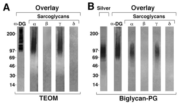
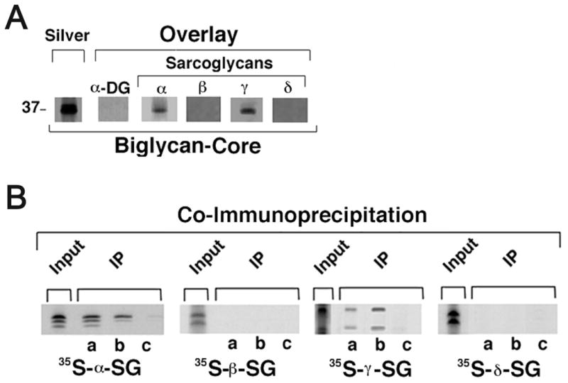
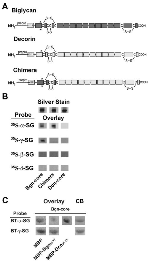
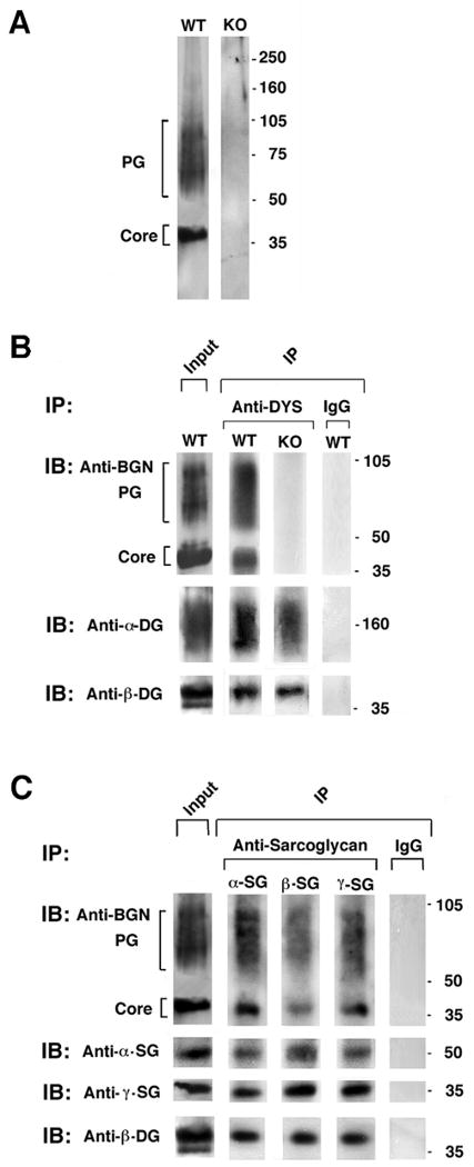
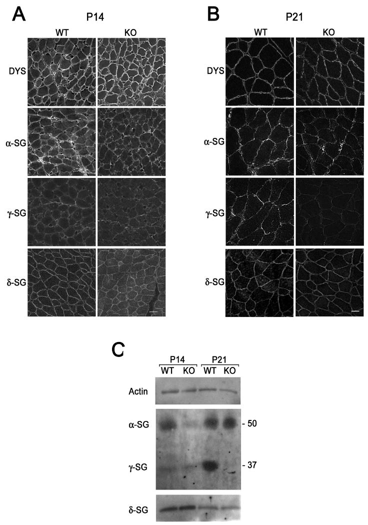
The dystrophin-associated protein complex (DAPC), which links the cytoskeleton to the extracellular matrix, is essential for muscle cell survival, and is defective in a wide range of muscular dystrophies. The DAPC contains two transmembrane subcomplexes-the dystroglycans and the sarcoglycans. Although several extracellular binding partners have been identified for the dystroglycans, none have been described for the sarcoglycan subcomplex. Here we show that the small leucine-rich repeat (LRR) proteoglycan biglycan binds to alpha- and gamma-sarcoglycan as judged by ligand blot overlay and co-immunoprecipitation assays. Our studies with biglycan-decorin chimeras show that alpha- and gamma-sarcoglycan bind to distinct sites on the polypeptide core of biglycan. Both biglycan proteoglycan as well as biglycan polypeptide lacking glycosaminoglycan (GAG) side chains are components of the dystrophin glycoprotein complex isolated from adult skeletal muscle membranes. Finally, our immunohistochemical and biochemical studies with biglycan null mice show that the expression of alpha- and gamma-sarcoglycan is selectively reduced in muscle from young (P14-P21) animals, while levels in adult muscle (> or = P35) are unchanged. We conclude that biglycan is a ligand for two members of the sarcoglycan complex and regulates their expression at discrete developmental ages.


Figures

Figure 1
Figure 1. Biglycan binds to α- and γ- sarcoglycan
A. Sarcoglycan binding to native biglycan. Postsynaptic membrane fractions from Torpedo electric organ (TEOM; 0.8 μg) were separated on SDS-PAGE gels, blotted onto nitrocellulose then probed with either 35S-methionine-labelled in vitro translated α-dystroglycan or sarcoglycans (α, β, γ, or δ) and analyzed by autoradiography. α-Dystroglycan as well as α- and γ-sarcoglycan bound to a polydisperse band whose center of migration was ∼125kD. In previous work a polypeptide with identical mobility, appearance and α–dystroglycan binding capacity was purified from these fractions and shown to be the proteoglycan biglycan (Bowe et al., 2000). No binding of β– or δ– sarcoglycan to this or any other polypeptide in these fractions was detected. B. Binding of α-dystroglycan and sarcoglycans to purified recombinant biglycan proteoglycan (Biglycan-PG). One microgram of biglycan was separated by SDS-PAGE and either stained with silver or blotted onto nitrocellulose (‘Overlay’) and probed as described above. α-Dystroglycan and α- and γ-sarcoglycan bind to this recombinant, GAG-containing biglycan proteoglycan, while no binding of β– or δ– sarcoglycan is detected.
Figure 2
Figure 2. The biglycan core polypeptide is sufficient for binding to both immobilized and soluble α- and γ-sarcoglycan
A. Purified recombinant biglycan core polypeptide (1μg) was separated by SDS-PAGE and either silver stained or blotted and probed as described above. α-Dystroglycan did not bind to this GAG-free biglycan. In contrast, both α- and γ- sarcoglycan bind to the biglycan core polypeptide. B. Co-immunoprecipitation of purified recombinant biglycan to recombinant sarcoglycans. His-tagged biglycan core polypeptide (0.5μg/ml) was incubated with the indicated 35S-methionine-labeled, in vitro translated sarcoglycan for 1 hr followed by either anti-biglycan (a), anti-poly-His (b) or normal rabbit Ig (c). Immune complexes were then precipitated with protein G beads and analyzed by SDS-PAGE and autoradiography. Note that both α- and γ- sarcoglycan co-immunoprecipitate with biglycan, while β- and δ- sarcoglycan do not. The labelling of the various sarcoglycans is shown by direct autoradiography of SDS-PAGE-separated in vitro translated polypeptides (‘Input’).
Figure 3
Figure 3. Distinct binding sites for α- and γ- sarcoglycan on the biglycan core polypeptide
A. Domain structure of biglycan, decorin and a biglycan-decorin chimera. The location of the pre-pro peptide (‘prepro’), 6-His tag, cysteine-rich amino- and carboxyl- domains, LRRs (ten open rectangles in the central domain; some schemes predict an 11th in the carboxyl-terminal cysteine-rich region) and GAG attachment sites (asterisks) are indicated. Note that these sites are present in the recombinant proteins used in this experiment, but they are not substituted with GAGs. B. Binding of sarcoglycans to biglycan, decorin and a chimera. One microgram of each of the purified recombinant proteins was separated by SDS-PAGE and either directly stained (‘silver’) or blotted and probed with 35S-methionine-labelled, in vitro-translated sarcoglycans as indicated. Both α- and γ- sarcoglycan bind to the immobilized biglycan core but not to decorin core. In contrast, only α-sarcoglycan binds to the biglycan-decorin chimeric protein. Thus the first 30 amino acids of biglycan is necessary for its binding to α-sarcoglycan. Neither β- nor δ- sarcoglycan bind to biglycan, decorin or the chimera. C. Competition studies. Sarcoglycan binding to purified recombinant biglycan core polypeptide in the presence of excess MBP-Bgn38-77 (amino acids 38 to 77 of biglycan) or MBP-Dcn31-71 (amino acids 31 to 71 of decorin). These sequences correspond to the first 40 amino acids of the mature biglycan and decorin polypeptides, respectively. Four micrograms of biglycan were separated by SDS-PAGE and either directly stained with Coomassie Blue (CB) or blotted and probed with biotinylated (BT), in vitro-translated α- or γ-sarcoglycan as indicated. Binding of α-sarcoglycan to full-length biglycan was inhibited in the presence of MBP-Bgn38-77, while binding between γ-sarcoglycan and biglycan binding remained unchanged. The decorin fusion protein did not inhibit this interaction.
Figure 4
Figure 4. Biglycan is a component of the native DAPC
A. Characterization of biglycan expressed in skeletal muscle membranes. Digitonin-solubilized, KCl-washed skeletal muscle membranes (3μg) from either wild type or biglycan null mice (KO; littermates, see methods) were separated on a 3-12% gradient SDS-PAGE. Western blotting for biglycan reveals two polypeptides, one migrating as a discrete band at ∼40kD (core) and a second polydisperse band with migration ranging from 60-100kD (PG). Both forms are absent in membranes prepared from biglycan null muscle. Identical results were obtained when intact membranes were used as starting material for the Western blot (not shown). B. Endogenous biglycan co-immunoprecipitates with dystrophin. Digitonin solubilized membranes were incubated with anti-dystrophin antisera DYS; or control IgG and immunoprecipitates IP were probed for dystroglycan (α and β) and biglycan by Western blotting. As expected, α– and β- dystroglycan co-immunoprecipitate with dystrophin. Notably, both the core and the proteoglycan forms of biglycan were present in these immune complexes. Neither biglycan form was detected in immunoprecipitates from biglycan null mice. C. Co-immunoprecipitation of biglycan with sarcoglycans. Digitonin-solubilized membranes were incubated with antibodies to α–, ß- or γ– sarcoglycan. Both the core and the proteoglycan forms of biglycan are detected in the immunoprecipitates. Equivalent levels of the three sarcoglycans are detected in the immunoprecipitates from wild type and biglycan null muscle membranes.
Figure 5
Figure 5. Reduced α- and γ- sarcoglycan expression in immature biglycan null mice
Immunohistochemical analysis of P14, A., and P21, B., mouse muscle. Sections of quadriceps femoris from congenic P14 wild type and biglycan null (KO) mice were sectioned, mounted on the same slides and immunolabelled for dystrophin or α-, γ-, or δ- sarcoglycan. A. α-Sarcoglycan levels at the sarcolemma of P14 biglycan null muscle are selectively reduced in the biglycan null as compared to wild type muscle. B. γ-Sarcoglycan expression is reduced in P21 biglycan null mice, while α-sarcoglycan is unchanged. The level of dystrophin and δ-sarcoglycan is the same at both ages. Scale bars = 10 μm. C. Biochemical analysis of sarcoglycan expression. α- and γ- Sarcoglycan expression is reduced at distinct postnatal ages in skeletal muscle membranes from immature mice. KCl-washed skeletal muscle membranes (3 μg) from P14 or P21 congenic wild type or biglycan null mice (KO) were separated via SDS-PAGE. Western blotting was performed for α-, γ-, or δ- sarcoglycan or actin (loading control). Western blotting for α- and γ- sarcoglycan was performed on the same gel. Western blotting for δ-sarcoglycan and actin was performed in parallel on the same gel. α- Sarcoglycan levels are reduced in P14 biglycan null membranes while γ-sarcoglycan levels are reduced in P21 biglycan null membranes. Equivalent levels of δ-sarcoglycan and actin expression are seen at both ages. Similar results were observed in muscles from two other sets of mice.

Similar articles

Cited by

References

    1. Adams ME, Butler MH, Dwyer TM, Peters MF, Murnane AA, Froehner SC. Two forms of mouse syntrophin, a 58 kd dystrophin-associated protein, differ in primary structure and tissue distribution. Neuron. 1993;11(3):531–540. - PubMed
    1. Adams ME, Kramarcy N, Fukuda T, Engel AG, Sealock R, Froehner SC. Structural abnormalities at neuromuscular synapses lacking multiple syntrophin isoforms. J Neurosci. 2004;24(46):10302–10309. - PMC - PubMed
    1. Ahn AH, Freener CA, Gussoni E, Yoshida M, Ozawa E, Kunkel LM. The three human syntrophin genes are expressed in diverse tissues, have distinct chromosomal locations, and each bind to dystrophin and its relatives. J Biol Chem. 1996;271(5):2724–2730. - PubMed
    1. Ameye L, Young MF. Mice deficient in small leucine-rich proteoglycans: novel in vivo models for osteoporosis, osteoarthritis, Ehlers-Danlos syndrome, muscular dystrophy, and corneal diseases. Glycobiology. 2002;12(9):107R–116R. - PubMed
    1. Bianco P, Fisher LW, Young MF, Termine JD, Robey PG. Expression and localization of the two small proteoglycans biglycan and decorin in developing human skeletal and non-skeletal tissues. J Histochem Cytochem. 1990;38(11):1549–1563. - PubMed

Publication types