

Restoring cerebral blood flow reveals neural regions critical for naming
- PMID: 16885220
- PMCID: PMC6673770
- DOI: 10.1523/JNEUROSCI.2088-06.2006
Restoring cerebral blood flow reveals neural regions critical for naming
Abstract
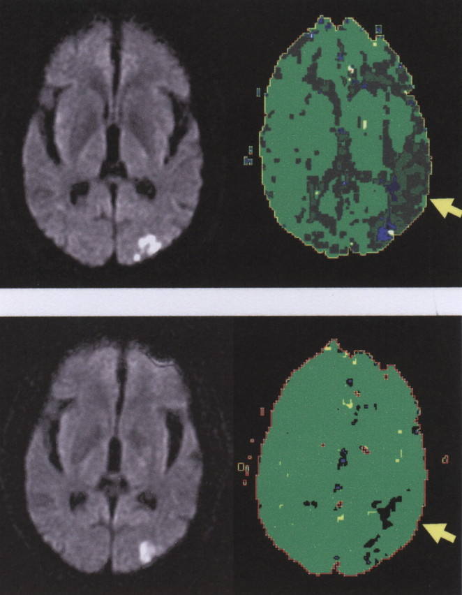
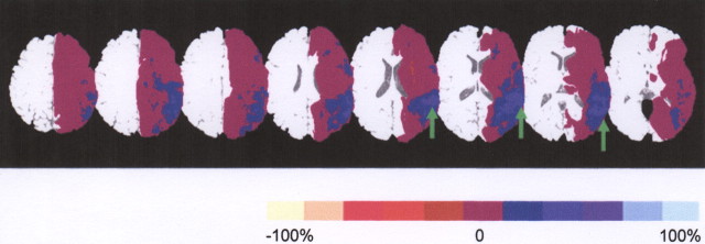
We identified areas of the brain that are critical for naming pictures of objects, using a new methodology for testing which components of a network of brain regions are essential for that task. We identified areas of hypoperfusion and structural damage with magnetic resonance perfusion- and diffusion-weighted imaging immediately after stroke in 87 individuals with impaired picture naming. These individuals were reimaged after 3-5 d, after a subset of patients underwent intervention to restore normal blood flow, to determine areas of the brain that had reperfused. We identified brain regions in which reperfusion was associated with improvement in picture naming. Restored blood flow to left posterior middle temporal/fusiform gyrus, Broca's area, and/or Wernicke's area accounted for most acute improvement after stroke. Results show that identifying areas of reperfusion that are associated with acute improvement of a function can reveal the brain regions essential for that function.
Figures
References
-
- Alexander MP, Naeser MA, Palumbo C (1990). Broca's area aphasias: aphasia after lesions including the frontal operculum. Neurology 40:353–362. - PubMed
-
- Blank SC, Scott SK, Murphy K, Warburton E, Wise RJ (2002). Speech production: Wernicke, Broca and beyond. Brain 125:1829–1838. - PubMed
-
- Broca P (1865). Sur la faculte du langage articule. Paris Bull Soc Anthr 6:337–393.
-
- Chatterjee A (2005). A madness to the methods in cognitive neuroscience? J Cogn Neurosci 17:847–849. - PubMed
-
- Croquelois A, Wintermark M, Reichhart M, Meuli R, Bogousslavsky J (2003). Aphasia in hyperacute stroke: language follows brain penumbra dynamics. Ann Neurol 54:321–329. - PubMed
Publication types
MeSH terms
Grants and funding
LinkOut - more resources
Full Text Sources
Medical