. 2006 Aug 2;26(31):8137-47.
doi: 10.1523/JNEUROSCI.1183-06.2006.

Actin-dependent activation of presynaptic silent synapses contributes to long-term synaptic plasticity in developing hippocampal neurons

Affiliations

Actin-dependent activation of presynaptic silent synapses contributes to long-term synaptic plasticity in developing hippocampal neurons

Jun Yao et al. J Neurosci. .

Abstract

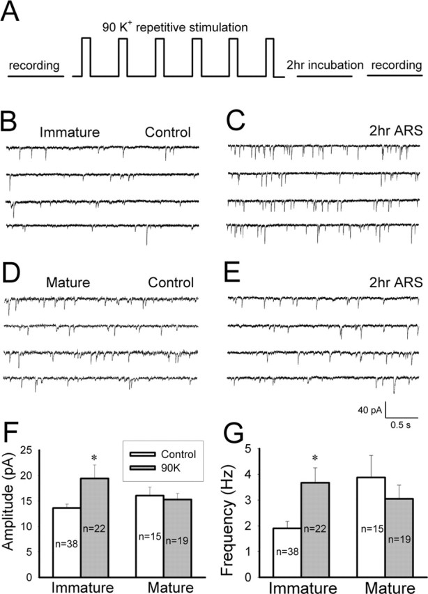
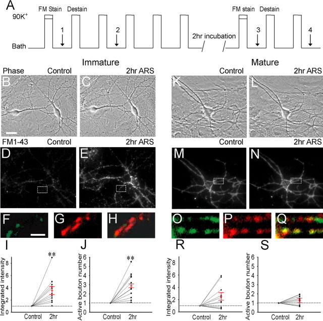
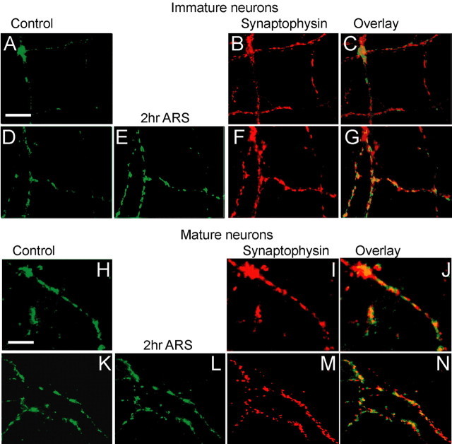
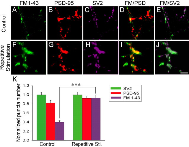
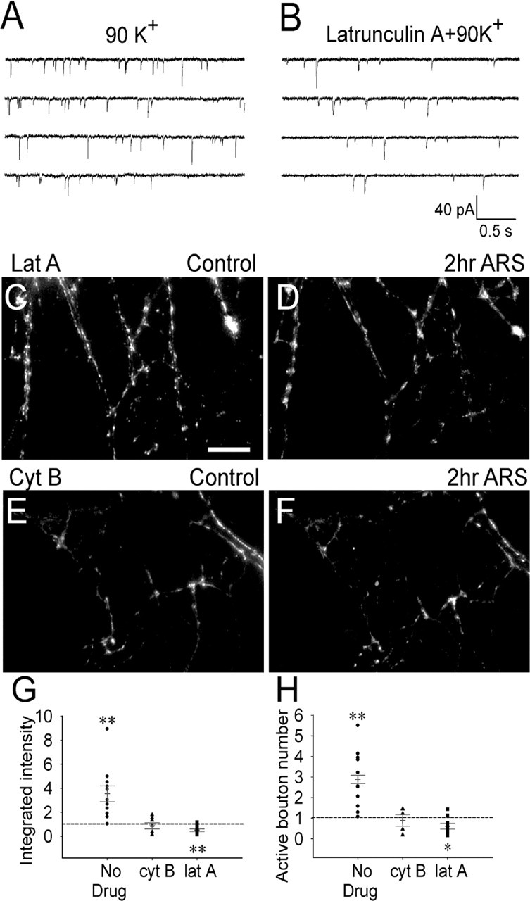
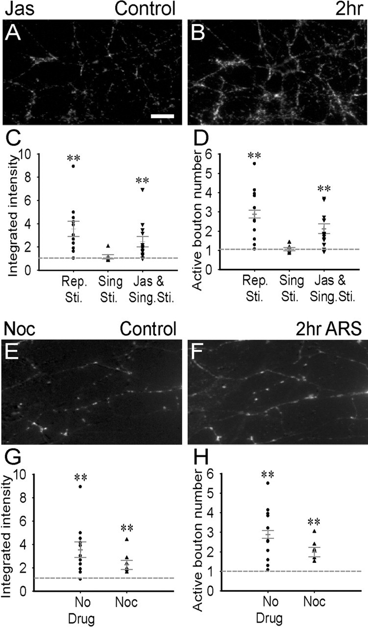
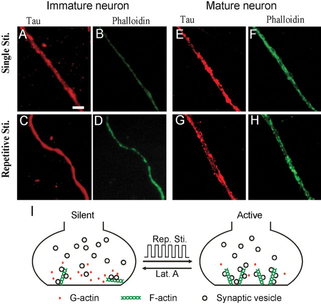
Developing neurons have greater capacity in experience-dependent plasticity than adult neurons but the molecular mechanism is not well understood. Here we report a developmentally regulated long-term synaptic plasticity through actin-dependent activation of presynaptic silent synapses in cultured hippocampal neurons. Live FM 1-43 imaging and retrospective immunocytochemistry revealed that many presynaptic boutons in immature neurons are functionally silent at resting conditions, but can be converted into active ones after repetitive neuronal stimulation. The activation of presynaptic silent synapses is dependent on L-type calcium channels and protein kinase A (PKA)/PKC signaling pathways. Moreover, blocking actin polymerization with latrunculin A and cytochalasin B abolishes long-term increase of presynaptic functional boutons induced by repetitive stimulation, whereas actin polymerizer jasplakinolide increases the number of active boutons in immature neurons. In mature neurons, however, presynaptic boutons are mostly functional and repetitive stimulation did not induce additional enhancement. Quantitative immunostaining with phalloidin revealed a significant increase in axonal F-actin level after repetitive stimulation in immature but not mature neurons. These results suggest that actin-dependent activation of presynaptic silent synapses contributes significantly to the long-term synaptic plasticity during neuronal development.


Figures

Figure 1.
Figure 1.
Repetitive stimulation induces long-term enhancement of synaptic transmission in immature but not mature hippocampal neurons. A, Diagram showing the experimental protocol. mEPSCs were recorded before and 2 h after six repetitive stimuli with 90 mm KCl solution. B, C, Representative traces illustrating mEPSCs recorded in immature neurons (7–11 d in culture) in the presence of TTX (0.5 μm) and BIC (20 μm) in control (B) and 2 h ARS (C). D, E, mEPSC traces in mature neurons (18–22 d in culture) in control (D) and 2 h after repetitive stimulation (E). F, Bar graphs showing that repetitive stimulation induced a significant increase in the average mEPSC amplitude in immature neurons (p < 0.003) but not mature neurons (p > 0.7). G, Bar graphs showing a significant increase in the mEPSC frequency in immature neurons (p < 0.03) but not mature neurons (p > 0.4) after repetitive stimulation. Error bars indicate SE. *p < 0.05.
Figure 2.
Figure 2.
Repetitive stimulation induces long-term enhancement of presynaptic function in immature but not mature neurons. A, Experimental protocol for FM 1-43 imaging and repetitive 90 K+ stimulation. Four fluorescence images were acquired (arrows, 1–4) for subtraction and subsequent imaging analysis. FMcontrol = FM1-stain − FM2-destain; FM2 h ARS = FM3-stain − FM4-destain. B, C, Phase images of the same immature neurons before (B) and 2 h after repetitive stimulation (C). Scale bar: (in B) B–E, K–N, 25 μm. D, E, Subtracted FM 1-43 images of the same neurons (corresponding to B and C) before (D) and 2 h after (E) repeated stimulation. F–H, Enlarged FM images from control (F, enlarged from D) and after stimulation (G, enlarged from E), as well as their merged picture (H). Scale bar: (in F) F–H, O–Q, 2.5 μm. I–J, Quantitative analysis showing a significant increase in the integrated FM intensity (I; n = 11; p < 0.01, paired Student’s t test) and FM-labeled active bouton number (J; n = 11; ∗∗p < 0.01) after repetitive stimulation in immature neurons. The FM intensity and active bouton number after repetitive stimulation were normalized to the control value unless otherwise stated. K, L, Phase images of the same mature neuron before (K) and 2 h after (L) repeated stimulation. M, N, Subtracted FM 1-43 images of the mature neuron before (M) and 2 h after (N) repeated stimulation. O–Q, Enlarged FM images from control (O, enlarged from M), after repetitive stimulation (P, enlarged from N), and merged picture (Q). R, S, Quantification of changes in the integrated FM intensity (R; n = 11; p > 0.1, paired t test) and the active bouton number (S; n = 11; p > 0.1) after repetitive stimulation in mature neurons. Error bars indicate SE.
Figure 3.
Figure 3.
Retrospective immunocytochemistry reveals presynaptic silent boutons in immature but not mature neurons. A, Activity-labeled FM 1-43 images in control immature neurons. B, Retrospective immunostaining of synaptophysin at the same field shown in A. C, Overlaid image of A and B showing that many synaptophysin-labeled boutons are not stained with FM 1-43. D, E, FM 1-43 images before (D) and 2 h after repetitive stimulation (E) in immature neurons. F, Retrospective immunostaining of synaptophysin at the same field shown in D and E. G, Overlaid image of E and F showing most of the synaptophysin-labeled boutons are now stained with FM 1-43 after repetitive stimulation. H, FM 1-43-labeled presynaptic boutons in control mature neurons. I, Synaptophysin-labeled presynaptic boutons. J, Overlaid image of H and I showing well correlated synaptophysin and FM puncta in mature neurons under the control condition. K, L, Presynaptic boutons labeled by FM 1-43 before (K) and 2 h after repetitive stimulation (L) in mature neurons. M, Synaptophysin-stained presynaptic boutons. N, Overlaid image of L and M. Scale bars, 15 μm.
Figure 4.
Figure 4.
Comparison of repetitive stimulation-induced changes of presynaptic versus postsynaptic puncta in immature neurons. A–C, Immature neurons under the control condition showing FM 1-43-labeled functional presynaptic boutons (A), PSD-95-labeled postsynaptic puncta (B), and SV2-labeled presynaptic puncta (C) in the same field. D, E, Overlaid images for FM 1-43 labeling together with PSD-95 staining (D), or with SV2 staining (E). F–H, Immature neurons after repetitive stimulation, illustrating FM 1-43-labeled functional presynaptic boutons (F), PSD-95-labeled postsynaptic puncta (G), and SV2-labeled presynaptic puncta (H). I, J, Overlaid images for FM 1-43 labeling with PSD-95 staining (I), or with SV2 staining (J). Scale bar, 10 μm. K, Quantification of changes of presynaptic versus postsynaptic puncta induced by repetitive stimulation. Data are normalized to the SV2-labeled puncta number. The FM 1-43-labeled active bouton number significantly increased from 40% in control (n = 12) to 90% after repetitive stimulation (n = 9; ***p < 0.001). Error bars indicate SE.
Figure 5.
Figure 5.
Dependence on L-type Ca2+ channels and PKA/PKC signaling pathways of the presynaptic enhancement. A, B, Typical mEPSC recordings in immature neurons under control and after repetitive stimulation in the presence of nimodipine (10 μm). Both the amplitude and frequency of mEPSCs did not show any significant change after nimodipine treatment (n = 12–14; p > 0.7). C, D, Subtracted FM 1-43 images of immature neurons before (C) and 2 h after repetitive stimulation in the presence of nimodipine (D). E, F, Subtracted FM 1-43 images of immature neurons before (E) and 2 h after repetitive stimulation (F) in the presence of PKA inhibitor H89 (1 μm). Scale bar: (in F) C–F, 15 μm. G, H, Quantitative analysis showing the effect of nimodipine, H89, and PKC inhibitor GF109203x (5 μm) on changes of presynaptic functional boutons after repetitive stimulation. Both nimodipine and H89 abolished the increase of the integrated FM intensity (G) and the active bouton number (H) after repetitive stimulation. GF109203x treatment decreased the fluorescence intensity (n = 7; p < 0.01) and the active bouton number (n = 7; p < 0.03) after repetitive stimulation, suggesting that PKC is important in maintaining normal synaptic functions. Error bars indicate SE. *p < 0.05; **p < 0.01.
Figure 6.
Figure 6.
Inhibition of actin polymerization abolishes presynaptic long-term enhancement in immature neurons. A, B, mEPSCs of immature neurons recorded after repetitive 90 K+ stimulation in the absence (A) or presence of latrunculin A (5 μm) (B). Latrunculin A treatment did not affect the amplitude of mEPSCs (n = 14–17; p > 0.1) but reduced the frequency significantly (p < 0.01). C, D, Subtracted FM 1-43 images of immature neurons before (C) and 2 h after (D) repeated stimulation with latrunculin A treatment. E, F, Subtracted FM 1-43 images of immature neurons before (E) and 2 h after (F) repeated stimulation with cytochalasin B (4 μm) treatment. Scale bar: (in C) C–F, 20 μm. G, H, Quantification of changes in the integrated fluorescence intensity (G) and active bouton number (H) after blocking actin polymerization. Cytochalasin B treatment abolished repetitive stimulation-induced increase of the integrated FM intensity (p > 0.13; n = 8) and the active bouton number (p > 0.16; n = 8), whereas latrunculin A significantly decreased the integrated FM intensity (**p < 0.01; n = 10) and the active bouton number (p < 0.03; n = 10). Error bars indicate SE. *p < 0.05.
Figure 7.
Figure 7.
Actin but not microtubule polymerization is critical to presynaptic long-term enhancement in immature neurons. A, B, Subtracted FM 1-43 images of immature neurons pretreated with actin polymerizer jasplakinolide (100 nm, 30 min) before (A) and 2 h after (B) single-spaced stimulation. C, D, Quantitative analysis showing a significant increase in the integrated FM intensity (C; p < 0.01; n = 13) and active bouton number (D; p < 0.01; n = 13) after jasplakinolide treatment. E, F, Subtracted FM 1-43 images of immature neurons pretreated with microtubule depolymerizer nocodazole (10 μm, 30 min) before (E) and 2 h after (F) repetitive stimulation. G, H, Quantitative analysis showing that after nocodazole treatment, repetitive stimulation still increased the integrated FM intensity (G; **p < 0.01; n = 8) and the active bouton number (H; p < 0.02; n = 8) in immature neurons. Scale bar: (in A) A, B, E, F, 20 μm. Error bars indicate SE.
Figure 8.
Figure 8.
Repetitive stimulation increases actin polymerization in immature but not mature neurons. A, B, Immature neurons coimmunostained with axon marker tau1 (A) and F-actin marker phalloidin (B) after single 90 K+ stimulation. C, D, Coimmunostaining of tau1 (C) and phalloidin (D) in immature neurons after repetitive stimulation. Repetitive stimulation induced a significant increase of phalloidin intensity in tau1-labeled axons of immature neurons (p < 0.001; n = 10). E, F, Mature neurons stained with tau1 (E) and phalloidin (F) after single stimulation. G, H, Mature neurons stained with tau1 (G) and phalloidin (H) after repetitive stimulation. Scale bar: (in A) A–H, 5 μm. No significant change in phalloidin intensity was found in mature axons after repetitive stimulation (p > 0.44; n = 10). I, Simplified model illustrating an important role of actin polymerization in the activation of presynaptic silent boutons induced by repetitive stimulation.

Similar articles

Cited by

References

    1. Abel T, Nguyen PV, Barad M, Deuel TA, Kandel ER, Bourtchouladze R (1997). Genetic demonstration of a role for PKA in the late phase of LTP and in hippocampus-based long-term memory. Cell 88:615–626. - PubMed
    1. Ackermann M, Matus A (2003). Activity-induced targeting of profilin and stabilization of dendritic spine morphology. Nat Neurosci 6:1194–1200. - PubMed
    1. Ahmari SE, Buchanan J, Smith SJ (2000). Assembly of presynaptic active zones from cytoplasmic transport packets. Nat Neurosci 3:445–451. - PubMed
    1. Allison DW, Gelfand VI, Spector I, Craig AM (1998). Role of actin in anchoring postsynaptic receptors in cultured hippocampal neurons: differential attachment of NMDA versus AMPA receptors. J Neurosci 18:2423–2436. - PMC - PubMed
    1. Allison DW, Chervin AS, Gelfand VI, Craig AM (2000). Postsynaptic scaffolds of excitatory and inhibitory synapses in hippocampal neurons: maintenance of core components independent of actin filaments and microtubules. J Neurosci 20:4545–4554. - PMC - PubMed

Publication types

LinkOut - more resources