. 2006 Aug;188(16):5865-77.
doi: 10.1128/JB.00060-06.

Characterization of the pilin ortholog of the Helicobacter pylori type IV cag pathogenicity apparatus, a surface-associated protein expressed during infection

Affiliations

Characterization of the pilin ortholog of the Helicobacter pylori type IV cag pathogenicity apparatus, a surface-associated protein expressed during infection

Joanna Andrzejewska et al. J Bacteriol. 2006 Aug.

Abstract

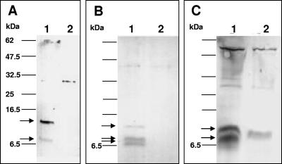
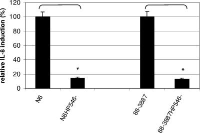
The Helicobacter pylori cag pathogenicity island (cag PAI) encodes components of a type IV secretion system (T4SS) involved in host interaction and pathogenicity. Previously, seven cag PAI proteins were identified as homologs of Agrobacterium tumefaciens Vir proteins, which form a paradigm T4SS. The T pilus composed of the processed VirB2 pilin is an external structural part of the A. tumefaciens T4SS. In H. pylori, cag-dependent assembly of pili has not been observed so far, nor has a pilin (VirB2) ortholog been characterized. We have here identified, using a motif-based search, an H. pylori cag island protein (HP0546) that possesses sequence and predicted structural similarities to VirB2-like pilins of other T4SSs. The HP0546 protein displays interstrain variability in its terminal domains. HP0546 was expressed as a FLAG-tagged fusion protein in Escherichia coli, A. tumefaciens, and H. pylori and was detected as either two or three bands of different molecular masses in the insoluble fraction, indicating protein processing. As reported previously, isogenic H. pylori mutants in the putative cag pilin gene had reduced abilities to induce cag PAI-dependent interleukin-8 secretion in gastric epithelial cells. Fractionation analysis of H. pylori, using a specific antiserum raised against an N-terminal HP0546 peptide, showed that the protein is partially surface exposed and that its surface localization depended upon an intact cag system. By immunoelectron microscopy, HP0546 was localized in surface appendages, with surface exposure of an N-terminal epitope. Pronounced strain-to-strain variability of this predicted surface-exposed part of HP0546 indicates a strong selective pressure for variation in vivo.


Figures

FIG. 1.
FIG. 1.
Alignment of H. pylori HP0546 protein (strain 26695) with various known type IV pilins of other bacteria. (A) The alignment shown was created with CLUSTAL W and revised manually (see Materials and Methods). Designations of protein origins, protein names, and bacterial species/plasmids are shown to the left of each alignment. Abbreviations for the plasmids/bacterial species: pKm10, pKM10 of E. coli; RP4, RP4 of E. coli; R388, R388 of E. coli; Eco, E. coli; atu, A. tumefaciens; bsui, Brucella suis; bhe, Bartonella henselae; borpe, Bordetella pertussis; Lepn, Legionella pneumoniae; Hpyl, H. pylori. Shading of the amino acids according to their physicochemical properties was performed with the GeneDoc software (www.psc.edu/biomed/genedoc). (B) The manual alignment of H. pylori HP0546 with the A. tumefaciens VirB2 type IV pilin protein shows 20% amino acid identity. The leader peptide cleavage site (putative for HP0546, at A29), the insertion site of an internal FLAG tag epitope (between V44 and I45) in HP0546, and the peptide (T31 to Q47; bold and underlined) which was used for generation of the specific antiserum are indicated in panel B.
FIG. 2.
FIG. 2.
Expression of FLAG-tagged HP0546 in three different bacterial hosts, E. coli, A. tumefaciens, and H. pylori, reveals that the protein is localized in the insoluble bacterial fractions and is processed. (A) Expression of HP0546 (of strain 26695) in E. coli MC1061. (B) Expression of HP0546 in A. tumefaciens NTREB1. (C) Expression of HP0546 in H. pylori 26695. Proteins (5 μg per lane) were separated on 16.5% Tris-Tricine SDS-PAGE gels, and Western blots were developed with anti-FLAG tag M2 monoclonal antibody. Only proteins from insoluble fractions after ultrasonication are shown, since no signals were detected in the soluble fractions. Lanes 1, insoluble fractions, which show HP0546 protein expression (plasmid constructs used for HP0546 expression are pCJ206 [A], pCJ207 [B], and pCJ205 [C]); lanes 2, insoluble fractions of negative controls without expression of specific FLAG-tagged proteins (plasmids used as negative controls are pCJ201-0 [A], pCJ203-1w [B], and pCJ201-0 [C]). Molecular masses of standard proteins are indicated on the left; arrows indicate proteins specifically detected with the anti-FLAG tag antibody.
FIG. 3.
FIG. 3.
Strain-specific detection of HP0546 protein in H. pylori using an antiserum raised against a peptide. Western blotting and immunolabeling of HP0546 (with and without FLAG tag), expressed in different H. pylori wild-type and plasmid-transformed strains and detected by a strain- and epitope-specific antiserum raised against an N-terminal peptide of HP0546, and control samples are shown. (A) Expression and strain-specific detection of HP0546 (from strain 26695) in E. coli MC1061 and in different H. pylori strains and mutants, including negative control samples which either do not express the protein (HP0546 mutant) or express a protein with a different N-terminal sequence (N6 and the N6 fliP mutant). Ten micrograms of protein (whole bacterial lysates) was loaded in each lane. (B) Western blot on which fractions of strain 88-3887 and its fliP (devoid of flagella), HP0546, and HP0544 mutants were analyzed, using the strain-specific anti-HP0546 antiserum. Insoluble (membrane) (I), soluble (cytoplasmic) (S), and surface-associated (extracellular) (E) bacterial fractions are depicted. HP0546 protein was found in the wild type in all three fractions, with predominance in the insoluble fraction. Note the slightly higher molecular mass of surface-associated HP0546. Four micrograms of protein was loaded in each lane. The upper two panels show fractionation controls for the same blot, developed using both anti-HPFlhA antiserum (raised against the membrane marker protein FlhA of the flagellar basal body) and anti-HPFlaB antiserum (raised against the extracellular marker protein flagellin B [20]). Arrows indicate protein bands, which were specifically detected by the respective antisera. Bands of lower molecular mass detected on FlhA-coincubated membranes and very weak bands detected by anti-HPFlaB are nonspecific bands.
FIG. 4.
FIG. 4.
Role of HP0546 in cell interaction. IL-8 release is reduced in HP0546 mutants compared to that in the isogenic wild-type 88-3887 (26695) and N6 strains. Human gastric epithelial cell lines (AGS for the 88-3887 strain pair and HM02 for the N6 strain pair) were coincubated for 20 h with the bacterial strains, and IL-8 secretion in the supernatants was measured by enzyme-linked immunosorbent assay. At least three independent cell infections were performed for each isogenic wild-type/mutant strain pair, and means/standard deviations for triplicate measurements of these triplicate infections are depicted. The results are summarized as relative IL-8 induction values (percentages of those for the respective wild-type strains, which were each set to 100%). Background values for mock-infected cells were deduced from all other values before the calculation. *, statistically significant differences in IL-8 induction between wild-type strains and isogenic mutants (paired t test; P < 0.005).
FIG. 5.
FIG. 5.
HP0546 is localized on bacterial surfaces using immunofluorescence microscopy of H. pylori specimens during cell infection, labeled with specific anti-HP0546 peptide antibody. (A) AGS cells infected with H. pylori 88-3887 wild-type bacteria for 4 h. (B) AGS cells coincubated with H. pylori 88-3887 HP0546 mutant bacteria for 4 h. Nonpermeabilized specimens were then stained by a lectin to visualize the AGS cell surface using wheat germ agglutinin coupled to Texas Red and subsequently fixed and immunolabeled using anti-HP0546 peptide antiserum (1:1,000), followed by anti-rabbit IgG coupled to Alexa Fluor 488 (1:5,000). Specimens were analyzed using confocal laser scanning microscopy, and representative focal planes are depicted as overlays between signals detected in the green (HP0546-specific signal) and red (cell surface staining) emission channels, combined with a differential interference contrast image of the same specimen. White bars are a size marker of 5 μm. No signal in the green channel (HP0546-specific) was detected in panel B.
FIG. 6.
FIG. 6.
Electron microscopy using anti-HP0546 antibody for immunolabeling of cag-positive H. pylori 88-3887 bacteria reveals surface-associated material. H. pylori bacteria were coincubated with AGS cells on electron microscopy grids for 2 h and then fixed and immunolabeled using gold-coupled secondary antibodies as described in Materials and Methods. Two representative bacterial specimens, which carry HP0546-specific material labeled with 10-nm gold grains at polar (top) and lateral (bottom) localizations, respectively, are shown. The bottom right panel depicts an inset at a higher magnification. Arrows point to flagella. Black bars are size markers of 0.5 μm.

References

    1. Akopyants, N. S., S. W. Clifton, D. Kersulyte, J. E. Crabtree, B. E. Youree, C. A. Reece, N. O. Bukanov, E. S. Drazek, B. A. Roe, and D. E. Berg. 1998. Analyses of the cag pathogenicity island of Helicobacter pylori. Mol. Microbiol. 28:37-53. - PubMed
    1. Alm, R. A., L.-S. L. Ling, D. T. Moir, B. L. King, E. D. Brown, P. C. Doig, D. R. Smith, B. Noonan, B. C. Guild, B. L. deJonge, G. Carmel, P. J. Tummino, A. Caruso, M. Uria-Nickelsen, D. M. Mills, C. Ives, R. Gibson, D. Merberg, S. D. Mills, Q. Jiang, D. E. Taylor, G. F. Vovis, and T. J. Trust. 1999. Genomic-sequence comparison of two unrelated isolates of the human gastric pathogen Helicobacter pylori. Nature 397:176-180. - PubMed
    1. Backert, S., E. Ziska, V. Brinkmann, U. Zimny-Arndt, A. Fauconnier, P. R. Jungblut, M. Naumann, and T. F. Meyer. 2000. Translocation of the Helicobacter pylori CagA protein in gastric epithelial cells by a type IV secretion apparatus. Cell. Microbiol. 2:155-164. - PubMed
    1. Brandt, S., T. Kwok, R. Hartig, W. Konig, and S. Backert. 2005. NF-kappaB activation and potentiation of proinflammatory responses by the Helicobacter pylori CagA protein. Proc. Natl. Acad. Sci. USA 102:9300-9305. - PMC - PubMed
    1. Buhrdorf, R., C. Forster, R. Haas, and W. Fischer. 2003. Topological analysis of a putative virB8 homologue essential for the cag type IV secretion system in Helicobacter pylori. Int. J. Med. Microbiol. 293:213-217. - PubMed

Publication types

MeSH terms

LinkOut - more resources