. 1990 Jan 25;265(3):1724-30.

Domain characterization of rabbit skeletal muscle myosin light chain kinase

Affiliations

Domain characterization of rabbit skeletal muscle myosin light chain kinase

B P Herring et al. J Biol Chem. .

Abstract

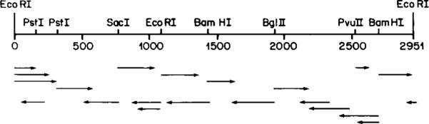
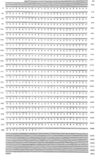
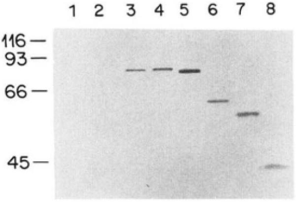
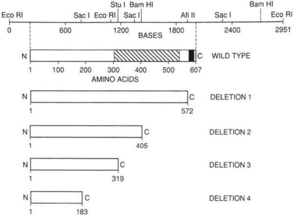
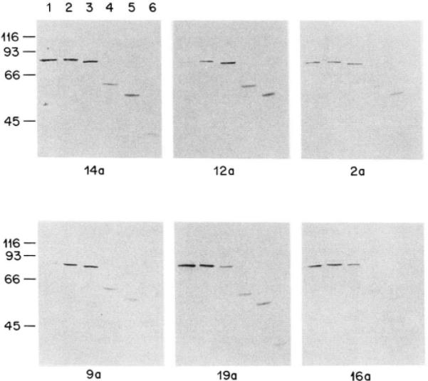
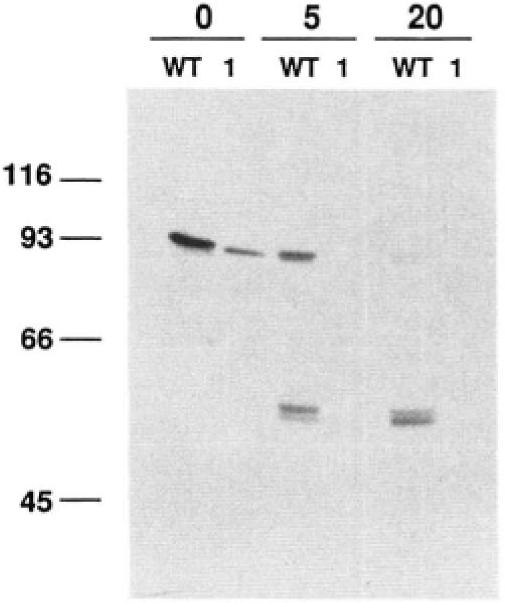
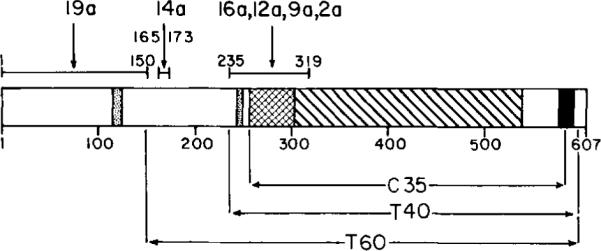
Myosin light chain kinase can be divided into three distinct structural domains, an amino-terminal "tail," of unknown function, a central catalytic core and a carboxy-terminal calmodulin-binding regulatory region. We have used a combination of deletion mutagenesis and monoclonal antibody epitope mapping to define these domains more closely. A 2.95-kilobase cDNA has been isolated that includes the entire coding sequence of rabbit skeletal muscle myosin light chain kinase (607 amino acids). This cDNA, expressed in COS cells encoded a Ca2+/calmodulin-dependent myosin light chain kinase with a specific activity similar to that of the enzyme purified from rabbit skeletal muscle. Serial carboxy-terminal deletions of the regulatory and catalytic domains were constructed and expressed in COS cells. The truncated kinases had no detectable myosin light chain kinase activity. Monoclonal antibodies which inhibit the activity of the enzyme competitively with respect to myosin light chain were found to bind between residues 235-319 and 165-173, amino-terminal of the previously defined catalytic core. Thus, residues that are either involved in substrate binding or in close proximity to a light chain binding site may be located more amino-terminal than the previously defined catalytic core.


Figures

Fig. 1
Fig. 1. Partial restriction map of the full length cDNA
Restriction endonuclease sites used in the sequencing strategy are indicated. Directional arrows indicate individual sequencing reactions.
Fig. 2
Fig. 2. Nucleotide sequence of the cDNA encoding rabbit skeletal muscle myosin light chain kinase
The nucleotide sequence of the full length cDNA and the deduced amino acid sequence of the coding region are shown. The internal EcoRI restriction site which was used to join the two halves of the clone is located at nucleotides 1096–1101. Amino acid residues are numbered on the left of the figure and nucleotide residues on the right.
Fig. 3
Fig. 3. Northern analysis of RNA isolated from several rabbit tissues
Total cellular RNAs (20 μg) from rabbit tissues were fractionated on a 1.2% agarose gel containing formaldehyde, transferred to nitrocellulose, and probed with a 32P-labeled cDNA probe. The cDNA used to probe the blot comprised residues 1–2848 of the full length cDNA. Following hybridization the blots were washed at high stringency (30 min at 68 °C in 0.2 × SSPE). Lane 1, fast twitch skeletal muscle RNA; lane 2, slow twitch skeletal muscle RNA; lane 3, uterus RNA; lane 4, liver RNA; lane 5, cardiac RNA. The positions of RNA molecular weight standards are shown in kilobases to the right side of the figure.
Fig. 4
Fig. 4. Immunoblot analysis of protein expressed from cDNAs transfected into COS cells
Wild type and truncated cDNAs were subcloned into the pCMV expression vector and transfected into COS cells as described under “Experimental Procedures.” The Nonidet P-40 lysates of the transfected cells were separated on 7.5% SDS-polyacrylamide gel electrophoresis, transferred to nitrocellulose, and reacted with polyclonal anti-skeletal muscle myosin light chain kinase as described under “Experimental Procedures.” Lane 1, lysate from cells with no DNA; lane 2, lysate from cells transfected with the full length cDNA in an antisense orientation; lane 3, 30 ng of purified rabbit skeletal muscle myosin light chain kinase; lane 4, lysate from cells transfected with the full length cDNA in the sense orientation; lanes 5–8, lysates from cells transfected with cDNA deletions 1–4, respectively. The positions of protein molecular weight standards are shown on the left side of the figure.
Fig. 5
Fig. 5. Partial restriction map of the full length cDNA and its alignment with the schematic linear amino acid sequences of the wild type and truncated myosin light chain kinases
Restriction sites used in the construction of the truncated cDNA are shown on the nucleotide map (top). The deduced amino acid residues of the wild type and truncated proteins are aligned below the nucleotide map. Each deletion has a common, wild type amino terminus; the carboxyl terminus of each expressed kinase is indicated in the figure. The proposed catalytic (hatched area) and calmodulin-binding (shaded) domains are indicated for the wild type kinase.
Fig. 6
Fig. 6. Immunoblot analysis of expressed proteins in COS cells with monoclonal antibodies
Immunoblots were prepared as described under “Experimental Procedures.” Each blot was reacted with the monoclonal antibody indicated in the figure. Antibody 19a was used at 11 μg/ml, 16a at 1 μg/ml, 14a at 5 μg/ml, 12a at 10 μg/ml, 9a at 5 μg/ml, and 2a at 4 μg/ml. The order of sample loading on each blot was identical. Lane 1, 30 ng of purified rabbit skeletal muscle myosin light chain kinase; lane 2, lysate from cells transfected with the full length cDNA in the sense orientation; lanes 3–6, lysates from cells transfected with deletions 1–4, respectively. The positions of protein molecular weight standards are indicated at the left side of the blots.
Fig. 7
Fig. 7. Tryptic digestion of myosin light chain kinase in COS cell lysates
COS cell lysates (30 μl) containing either wild type kinase (WT) or deletion mutant 1 (1) were digested with 1 μg of trypsin for 0, 5, or 20 min at 23 °C. The digestion was stopped by boiling the samples in five volumes of 10% SDS, 0.1 M dithiothreitol, 125 mM Tris at pH 6.8 and 20% sucrose. The partially proteolyzed fragments were subsequently analyzed by immunoblot using polyclonal anti-myosin light chain kinase antibodies.
Fig. 8
Fig. 8. Summary of epitope maps and domain organization of rabbit skeletal muscle myosin light chain kinase
This schematic diagram depicts the location of the functional domains of skeletal muscle myosin light chain kinase. The calmodulin-binding domain (Edelman et al., 1985; Blumenthal et al., 1985) is shaded, the catalytic core defined by homology with other protein kinases (Hanks et al., 1988) is hatched, whereas the catalytic domain extended to include a putative light chain-binding site defined by our studies is cross-hatched. The location of the epitopes of each monoclonal antibody are indicated. The stippled area represents potential sites of high antigenicity as predicted from the hydrophilic profile of the amino acid sequence.

References

    1. Andersson S, Davis DN, Dahlbäck H, Jörnvall H, Russell DW. J. Biol. Chem. 1989;264:8222–8229. - PubMed
    1. Blumenthal DK, Stull JT. Biochemistry. 1980;19:5608–5614. - PubMed
    1. Blumenthal DK, Stull JT. Biochemistry. 1982;21:2386–2391. - PubMed
    1. Blumenthal DK, Takio K, Edelman AM, Charbonneau H, Titani K, Walsh KA, Krebs EG. Proc. Natl. Acad. Sci. U. S. A. 1985;82:3187–3191. - PMC - PubMed
    1. Edelman AM, Takio K, Blumenthal DK, Hansen RS, Walsh KA, Titani K, Krebs EG. J. Biol. Chem. 1985;260:11275–11285. - PubMed

Publication types

Associated data