. 2006 Aug;116(8):2094-104.
doi: 10.1172/JCI27179.

Pathogenic role for skin macrophages in a mouse model of keratinocyte-induced psoriasis-like skin inflammation

Affiliations

Pathogenic role for skin macrophages in a mouse model of keratinocyte-induced psoriasis-like skin inflammation

Athanasios Stratis et al. J Clin Invest. 2006 Aug.

Abstract

Psoriasis is a common skin disease, the pathogenesis of which has not yet been resolved. In mice, epidermis-specific deletion of inhibitor of NF-kappaB (IkappaB) kinase 2 (IKK2) results in a skin phenotype that mimics human psoriasis in several aspects. Like psoriasis, this skin disease shows pronounced improvement when mice are treated with a TNF-neutralizing agent. We have found previously that this phenotype does not depend on the presence of alphabeta T lymphocytes. In order to evaluate contributions of other immune cell populations to the skin disease, we selectively eliminated macrophages and granulocytes from the skin of mice with epidermis-specific deletion of IKK2 (K14-Cre-IKK2fl/fl mice). Elimination of skin macrophages by subcutaneous injection of clodronate liposomes was accompanied by inhibition of granulocyte migration into the skin and resulted in a dramatic attenuation of psoriasis-like skin changes. The hyperproliferative, inflammatory skin disease in K14-Cre-IKK2fl/fl mice was a direct consequence of the presence of macrophages in the skin, as targeted deletion of CD18, which prevented accumulation of granulocytes but not macrophages, did not lead to major changes in the phenotype. Targeted deletion of the receptor for IFN-gamma revealed that the pathogenesis of the skin disease does not depend on classical IFN-gamma-mediated macrophage activation. Our results demonstrate that in mice epidermal keratinocytes can initiate a hyperproliferative, inflammatory, IFN-gamma-independent, psoriasis-like skin disease whose development requires essential contributions from skin macrophages but not from granulocytes or alphabeta T lymphocytes.


Figures
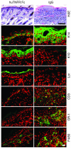
Figure 1
Figure 1. Improvement of psoriasis-like skin disease in K14-Cre-IKK2fl/fl mice by administration of huTNFR:Fc.
Light microscopic and confocal images of paraffin-embedded skin sections (H&E, K14, K10, and loricrin [Lor]) and confocal images of cryostat skin sections (CD3, GR-1, F4/80) obtained at P7 from K14-Cre-IKK2fl/fl mice injected with 20 μg per mouse of huTNFR:Fc (left panels) or human IgG (right panels). Top 2 panels are stained with H&E. For immunostainings, indicated markers are stained green, and nuclei are stained red. Scale bar: 100 μm (H&E); 40 μm (immunostaining).
Figure 2
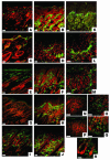
Figure 2. Characterization of macrophages and dendritic cells in the skin of K14-Cre-IKK2fl/fl mice.
Confocal images of immunostainings of skin of untreated (B, D, F, H, J, KM, P, and Q) or clodronate liposome–treated (R) K14-Cre-IKK2fl/fl mice and of control mice (A, C, E, G, I, N, and O) with antibodies against CD11b (A and B), CD14 (C and D), CD206 (E and F), CD83 (G and H), CD16/32 (I and J), and F4/80 (L and M). Respective markers are stained green; nuclei are stained red. C and D show unspecific staining of sebaceous glands due to the use of streptavidin-coupled fluorochrome for detection of biotinylated primary antibodies. (K) Double staining with antibodies against CD83 (green) and CD80 (red). (L and M) F4/80-positive epithelium-lining macrophages in a developing lesion at P4 (L) and in a fully developed lesion at P7 (M). (NR) Staining with mPDCA-1 antibody (green). Nuclei are stained red. N and P show upper dermis; O, Q, and R show deep dermis and subcutis. Scale bars: 40 μm (AM); 80 μm (NR).
Figure 3
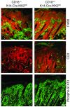
Figure 3. Improvement of the psoriasis-like skin phenotype after injection of clodronate liposomes.
Light microscopic images of paraffin-embedded skin sections (top 3 panels) and confocal images of cryostat skin sections (bottom 6 panels) from control mice and K14-Cre-IKK2fl/fl mice with clodronate or control liposomes injected as indicated. Control mice were not injected. Sections in the upper panels are stained H&E. Immunostainings in the middle and lower panels are against F4/80 and phosphorylated STAT3 (Phospho-STAT3), respectively (green). Nuclei are shown in red. White dotted line indicates the position of the epidermal basement membrane. Scale bar: 100 mm (H&E); 40 μm (immunostaining).
Figure 4
Figure 4. Injection of clodronate liposomes normalizes epidermal differentiation and prevents migration of immune cells.
Immunostainings of paraffin-embedded skin sections with antibodies against the epidermal differentiation markers K14, K10, loricrin, and filaggrin (Fil) or of frozen skin with antibodies against the immune cell markers GR-1 for granulocytes and CD3 for T lymphocytes. Skin samples were obtained at P7 from K14-Cre-IKK2fl/fl mice injected with control liposomes (left panels), clodronate liposomes (center panels), from uninjected control mice (right panels). Indicated markers are stained green, and nuclei are stained red. Scale bar: 40 μm.
Figure 5
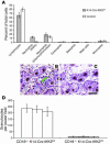
Figure 5. Targeted deletion of CD18 prevents granulocyte migration into the skin of K14-Cre-IKK2fl/fl mice.
(A) Results of white blood cell counting from blood smears of 3 K14-Cre-IKK2fl/fl mice and 2 control mice. Note relative increase in numbers of neutrophils (stabs and polymorphonuclear granulocytes) and relative decrease in numbers of lymphocytes. (B and C) Light microscopic images of chloroacetate esterase–stained, paraffin-embedded skin sections from CD18+/– K14-Cre-IKK2fl/fl mice (B) and CD18–/– K14-Cre-IKK2fl/fl mice (C). Granulocytes are stained red (arrow). Scale bar: 50 μm. (D) Results of counting of chloroacetate esterase–positive granulocytes in skin sections from 8 CD18–/– K14-Cre-IKK2fl/fl mice and 6 CD18+/+ or CD18+/– K14-Cre-IKK2fl/fl mice. Bars represent means of 10 fields for each mouse ± SD.
Figure 6
Figure 6. Elimination of granulocytes from the skin of K14-Cre-IKK2fl/fl mice does not prevent the development of the psoriasis-like skin disease.
Light microscopical (top 4 panels) and confocal images (bottom 12 panels) of paraffin-embedded skin sections obtained from mice of the indicated genotypes at P7. For immunostainings, indicated markers are stained green, and nuclei are stained red. Scale bars: 100 μm (H&E); 40 μm (immunostaining).
Figure 7
Figure 7. Elimination of granulocytes from the skin of K14-Cre-IKK2fl/fl mice does not suppress inflammation.
Confocal images of frozen skin sections from CD18–/– K14-Cre-IKK2fl/fl mice (left panels) and CD18+/– K14-Cre-IKK2fl/fl mice (right panels) immunostained for the indicated immune cell markers and phosphorylated STAT3 (green). Nuclei are stained red. White dotted lines indicate the position of the epidermal basement membrane. Scale bar: 40 μm.
Figure 8
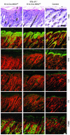
Figure 8. Targeted deletion of IFN-γ receptor does not prevent the development of the psoriasis-like skin disease in K14-Cre-IKK2fl/fl mice.
Light microscopic and confocal images of paraffin-embedded skin sections (H&E, K14, K10, loricrin, and filaggrin) and confocal images of cryostat skin sections (F4/80) obtained at P7 from K14-Cre-IKK2fl/fl mice, IFN-γR–/– K14-Cre-IKK2fl/fl mice, and control mice. Top 3 panel are stained with H&E. For immunostainings, indicated markers are stained green, and nuclei are stained red. Scale bar: 100 μm (H&E); 40 μm (immunostaining).

Comment in

References

    1. Lever, W.F., and Schaumburg-Lever, G. 1983. Histopathology of the skin. J.B. Lippincott Company. Philadelphia, Pennsylvania, USA. 848 pp.
    1. Schön M., et al. Critical role of neutrophils for the generation of psoriais skin lesions in flaky skin mice. J. Invest. Dermatol. 2000;114:976–983. - PubMed
    1. Schön M., Behmenburg D., Denzer D., Schön M.P. Pathogenic function of IL-1 beta in psoriasiform skin lesions of flaky skin (fsn/fsn) mice. Clin. Exp. Immunol. 2001;123:505–510. - PMC - PubMed
    1. Schön M.P., Detmar M., Parker C.M. Murine psoriasis-like disorder induced by naive CD4+ T-cells. Nat. Med. 1997;3:183–188. - PubMed
    1. Blessing M., Schirmacher P., Kaiser S. Overexpression of bone morphogenetic protein-6 (BMP-6) in the epidermis of transgenic mice: inhibition or stimulation of proliferation depending on the pattern of transgene expression and formation of psoriatic lesions. J. Cell Biol. 1996;135:227–239. - PMC - PubMed

Publication types

MeSH terms