. 2006 Aug;116(8):2105-14.
doi: 10.1172/JCI27180.

Activated macrophages are essential in a murine model for T cell-mediated chronic psoriasiform skin inflammation

Affiliations

Activated macrophages are essential in a murine model for T cell-mediated chronic psoriasiform skin inflammation

Honglin Wang et al. J Clin Invest. 2006 Aug.

Abstract

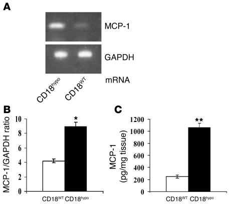
The CD18 hypomorphic (CD18hypo) PL/J mouse model clinically resembling human psoriasis is characterized by reduced expression of the common chain of beta2 integrins (CD11/CD18) to only 2-16% of WT levels. Previously we found that this chronic psoriasiform skin inflammation also depends on the presence of CD4+ T cells. Herein we investigated the role of macrophages in this CD18hypo mouse model. Activated macrophages were significantly increased in lesional skin as well as in inflamed skin draining lymph nodes (DLNs) of affected CD18hypo mice and were identified as being an important source of TNF-alpha in vivo. Both depletion of macrophages and neutralization of TNF-alpha resulted in a significant alleviation of psoriasiform skin inflammation. As monocyte chemotactic protein 1 was enhanced in lesional skin of affected CD18hypo mice, we intradermally injected recombinant murine monocyte chemotactic protein-1 (rJE/MCP-1) alone or in combination with rTNF-alpha into the skin of healthy CD18hypo mice. Only simultaneous injection of rJE/MCP-1 and rTNF-alpha, but neither substance alone, resulted in the induction of psoriasiform skin inflammation around the injection sites with recruitment and activation of macrophages. Collectively, our data suggest that maintenance of psoriasiform skin inflammation critically depends on efficient recruitment and activation of macrophages with sufficient release of TNF-alpha.


Figures

Figure 1
Figure 1. Increase in macrophages numbers in lesional skin and skin DLNs of affectedCD18hypo mice.
(A and B) Skin cryosections from CD18WT (A) and affected CD18hypo mice (B) were stained with F4/80–Alexa 488 for infiltrating macrophages (green) into the skin. Cell nuclei (blue) were counterstained with DAPI. e, epidermis; d, dermis; h, hair follicle. Dotted lines indicate the border between epidermis and dermis. Original magnification, ×40; inset, ×100. (C) To quantify macrophages in the skin of affected CD18hypo and CD18WT mice, the positively stained cells were calculated. For all measurements, the median of macrophages counted in 12 high-power fields (HPF) is presented (n = 4). ##P < 0.0001, Student’s t test. (D and E) Immunostaining with macrophage/monocyte-FITC (clone MOMA-2) was performed on cryosections of skin DLNs from CD18WT (D) and affected CD18hypo mice (E). Infiltrated macrophages (green) were found in the medullar and subcapsular sinuses, as indicated by arrows. Cell nuclei (red) were counterstained with propidium iodide (PI). Original magnification, ×20. (F and G) To quantify macrophages in the skin DLNs of CD18WT (F) and affected CD18hypo mice (G), skin DLN cells were labeled with MOMA-2–FITC and analyzed by flow cytometry. Dotted line, isotype control; gray histogram, MOMA-2 staining. (H) Total number of macrophages in skin DLNs of CD18hypo and CD18WT mice (n = 6). One representative experiment of 3 is shown. **P < 0.01, Student’s t test.
Figure 2
Figure 2. Activated macrophages are an important source of TNF-α in the lesional skin of affectedCD18hypo mice.
Double immunostaining with anti-mouse TNF-α and F4/80 mAb was performed on cryosections derived from the lesional skin of affected CD18hypo mice. (A) Macrophages (green) stained with F4/80–Alexa 488. (B) TNF-α (red) stained with TNF-α–Cy3. (C) Cell nuclei were stained with DAPI (blue). (D) Overlay depicting double staining of TNF-α and macrophages (yellow). (DG) Both classically and alternatively activated macrophages were present in the lesional skin of CD18hypo mice. To characterize the activation pattern of macrophages infiltrating the skin of affected CD18hypo mice, cryosections from lesional skin were double stained with F4/80–Alexa 488 (green) and the markers of classically activated macrophages TNF-α–Cy3 (red) (D) and iNOS-Cy3 (red) (E) or with the markers of alternatively activated macrophages Dectin 1–Cy3 (red) (F) and Arginase 1–Cy3 (G). Overlay (yellow) represents double-positive cells, i.e., macrophages bearing specific activation markers. Cell nuclei were counterstained with DAPI (blue). Dotted lines indicate the border between epidermis and dermis. Original magnification, ×20.
Figure 3
Figure 3. Reduction of the psoriasiform phenotype of affectedCD18hypo mice by administration of etanercept.
(A and B) A significant difference in the adapted PASI score appeared after treatment with etanercept (A; n = 5; #P = 0.0079, Student’s t test), but not after treatment with 0.9% NaCl control (B; n = 3; P = 0.6514, Student’s t test). (CE) Significantly reduced numbers of macrophages in skin DLNs of affected CD18hypo mice treated with etanercept (C) were observed after 30 days compared with controls (D and E). (FH) Decreased numbers of MHCII+ cells in skin DLNs of affected CD18hypo mice treated with etanercept (F) were detected after 30 days compared with controls (G and H). M1, MHCII cells; M2, MHCII+ cells. **P < 0.01, Student’s t test. (I and J) Decreased expression of TNF-α (red) was observed in the skin of affected CD18hypo mice treated with etanercept after 30 days (I) compared with controls (J). Cell nuclei were stained with DAPI (blue). (K and L) Reduced numbers of F4/80+ macrophages (green) were observed in the skin of affected CD18hypo mice treated with etanercept after 30 days (K) compared with controls (L). Cell nuclei (red) were counterstained with propidium iodide. Original magnification, ×20.
Figure 4
Figure 4. Improvement of the psoriasiform phenotype of affectedCD18hypo mice by depletion of macrophages after local injection with clodronate liposomes.
Clodronate liposomes were injected subcutaneously (50 mg/200 μl, administered weekly). (A and B) Representative clinical picture of a CD18hypo mouse with severe psoriasiform dermatitis before (A) and after 40 days of treatment with clodronate liposomes (B). (C and D) The severity of the psoriasiform phenotype was significantly reduced after clodronate treatment (C; n = 6) but not after treatment with control liposomes (D; n = 4; P = 0.3429, Student’s t test). **P < 0.01. (EH) Efficiency of depletion was evaluated by immunostaining. Clodronate liposome treatment dramatically reduced the number of F4/80+ macrophages (green) in injected skin areas (F) and skin DLNs (H) of CD18hypo mice compared with skin (E) and skin DLNs (G) from mice treated with control liposomes. Depletion of inflammatory macrophages also caused a decrease in TNF-α expression (red): skin samples from CD18hypo mice treated with control liposomes showed strong TNF-α expression (I), whereas expression in clodronate liposome–treated skin was virtually undetectable (J). GR-1 staining (red) of skin from CD18hypo mice 40 days after clodronate liposome injection (K), when skin inflammation and phenotype had completely resolved, revealed that granulocytes were removed, whereas GR-1+ granulocytes were still present after PBS-liposome treatment (L). Dotted lines indicate the border between epidermis and dermis. Cell nuclei were counterstained with propidium iodine (red) or DAPI (blue) as indicated. Original magnification, ×40 (E, F, K, and L); ×10 (G and H).
Figure 5
Figure 5. Enhanced MCP-1 mRNA and protein levels in the lesional skin of affectedCD18hypo mice.
Total RNA was extracted from skin of affected CD18hypo and CD18WT mice (n = 3). cDNA was analyzed for MCP-1 and GAPDH mRNA expression by semiquantitative RT-PCR. (A) Specific bands for MCP-1 (142 bp) and internal control GAPDH were visualized by ethidium bromide in 1% agarose gels. (B and C) Both MCP-1 mRNA levels as determined by quantitative real-time RT-PCR (B) and MCP-1 protein levels as determined by specific MCP-1 ELISA (C) were increased in lesional skin of affected CD18hypo mice compared with CD18WT controls. MCP-1 protein levels are expressed in relation to tissue weight as the mean ± SD for triplicate samples of each mouse. *P < 0.05; **P < 0.01.
Figure 6
Figure 6. The injection of a combination of rJE/MCP-1 and rTNF-α induces psoriasiform inflammatory skin lesions inCD18hypo PL/J mice.
Neither injection of rJE/MCP-1 nor rTNF-α alone resulted in psoriasiform skin disease (Supplemental Figure 3 and data not shown). (A and B) A combination of 0.2 μg murine rJE/MCP-1 and 5,000 U rTNF-α, injected in 200 μl PBS intradermally in the upper back skin of nonaffected CD18hypo PL/J mice (A), led to the appearance of erythematous plaques covered with scales and crusts starting at day 4 after administration (B). (C and D) Skin samples from paraffin sections were stained with H&E before (C) and 10 days after treatment, when epidermal hyperplasia and an abundant inflammatory infiltrate were observed within the dermis (D). (E and F) Cryosections from skin samples before (E) and after treatment stained for the keratinocyte marker K14–Alexa 488 (green) showed an increase in epidermal thickness and enhanced expression of the K14 proliferation-associated marker 10 days after treatment (F). (G and H) At this same time point, a dermal inflammatory infiltrate predominantly consisting of F4/80–Alexa 488+– (green) and TNF-α–PE+–activated (red) macrophages (overlay, yellow) was identified (H) compared with day 0 (G). (I and J) Interestingly, virtually no CD4-FITC+ T cells (green) were present in the dermis before (I) or after treatment with rMCP-1 and rTNF-α (J). Cell nuclei were counterstained with DAPI (blue). Dotted lines indicate the border between epidermis and dermis. Original magnification, ×20.

Comment in

References

    1. Nickoloff B.J. The immunologic and genetic basis of psoriasis. Arch. Dermatol. 1999;135:1104–1110. - PubMed
    1. Nickoloff B.J., Nestle F.O. Recent insights into the immunopathogenesis of psoriasis provide new therapeutic opportunities. J. Clin. Invest. 2004;113:1664–1675. doi: 10.1172/JCI200422147. - DOI - PMC - PubMed
    1. Gillitzer R., et al. MCP-1 mRNA expression in basal keratinocytes of psoriatic lesions. J. Invest. Dermatol. 1993;101:127–131. - PubMed
    1. van den Oord J.J., de Wolf-Peeters C. Epithelium-lining macrophages in psoriasis. Br. J. Dermatol. 1994;130:589–594. - PubMed
    1. Djemadji-Oudjiel N., Goerdt S., Kodelja V., Schmuth M., Orfanos C.E. Immunohistochemical identification of type II alternatively activated dendritic macrophages (RM 3/1+3, MS-1+/–, 25F9-) in psoriatic dermis. Arch. Dermatol. Res. 1996;288:757–764. - PubMed

Publication types

MeSH terms