. 2006 Aug;116(8):2161-72.
doi: 10.1172/JCI26185.

Autophagy is involved in T cell death after binding of HIV-1 envelope proteins to CXCR4

Affiliations

Autophagy is involved in T cell death after binding of HIV-1 envelope proteins to CXCR4

Lucile Espert et al. J Clin Invest. 2006 Aug.

Abstract

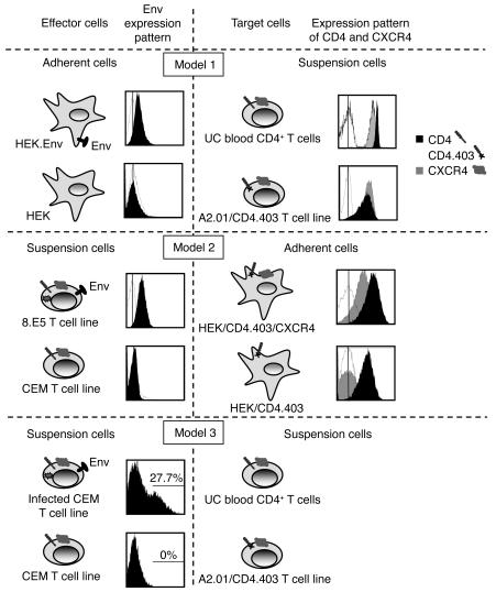
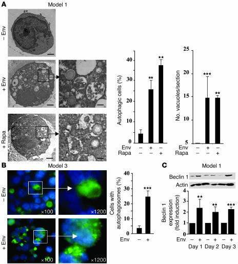
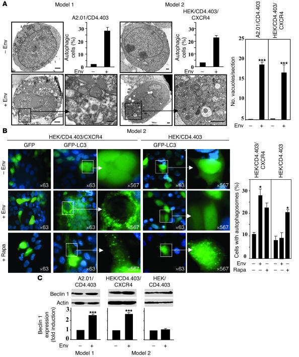
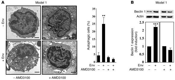
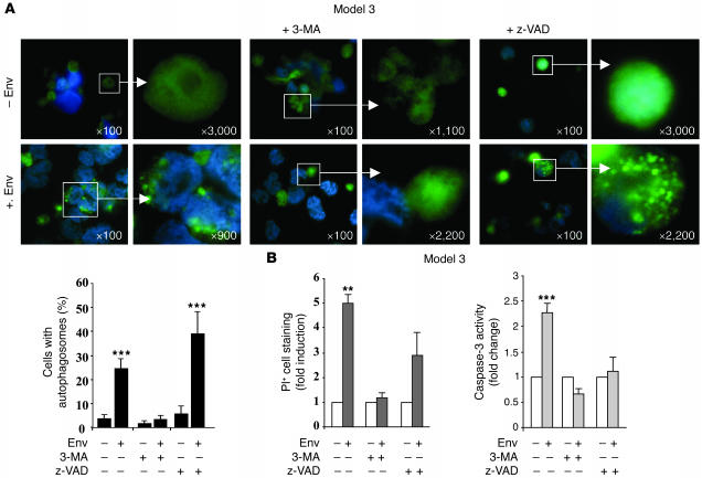
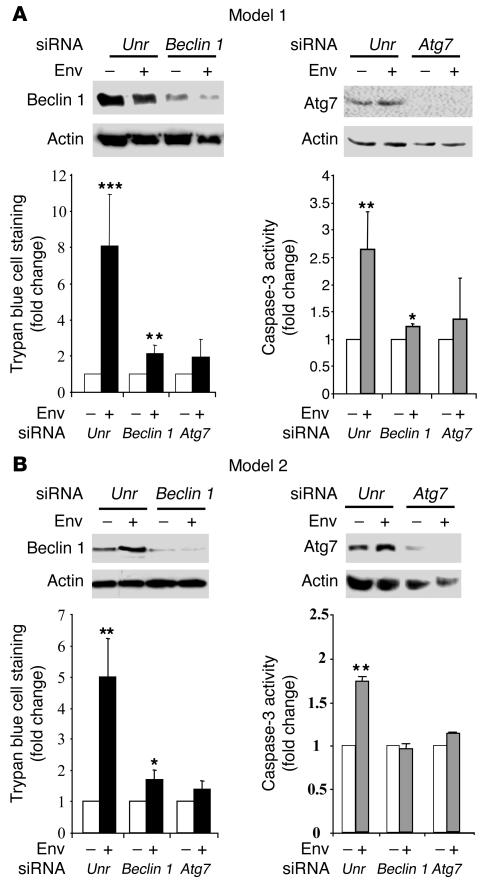
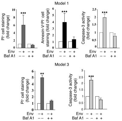
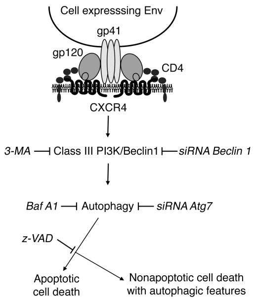
HIV-1 envelope glycoproteins (Env), expressed at the cell surface, induce apoptosis of uninfected CD4+ T cells, contributing to the development of AIDS. Here we demonstrate that, independently of HIV replication, transfected or HIV-infected cells that express Env induced autophagy and accumulation of Beclin 1 in uninfected CD4+ T lymphocytes via CXCR4. The same phenomena occurred in a T cell line and in transfected HEK.293 cells that expressed both wild-type CXCR4 and a truncated form of CD4 that is unable to bind the lymphocyte-specific protein kinase Lck. Env-mediated autophagy is required to trigger CD4+ T cell apoptosis since blockade of autophagy at different steps, by either drugs (3-methyladenine and bafilomycin A1) or siRNAs specific for Beclin 1/Atg6 and Atg7 genes, totally inhibited the apoptotic process. Furthermore, CD4+ T cells still underwent Env-mediated cell death with autophagic features when apoptosis was inhibited. These results suggest that HIV-infected cells can induce autophagy in bystander CD4+ T lymphocytes through contact of Env with CXCR4, leading to apoptotic cell death, a mechanism most likely contributing to immunodeficiency.


Figures

Figure 1
Figure 1. Description of the cellular models.
In model 1, target CD4+ T cells (UC blood CD4+ T cells and a T cell line expressing CXCR4 and CD4.403) are cocultured with effector HEK cells with or without Env expression. In model 2, HEK/CD4.403/CXCR4 and HEK/CD4.403 cells are cocultured with effector T cells with or without Env expression. In model 3, target CD4+ T cells are cocultured with HIVNL4-3-infected or uninfected T cells. Expression of gp120 at the surface of HEK.Env, 8.E5, and HIVNL4-3-infected effector cells as well as parental HEK and CEM cells — used as negative controls — were analyzed by flow cytometry after incubation of the cells with PBS (white histograms) or with PBS containing anti-gp120 human polyclonal Ab (black histograms). Bound Ab was detected with a secondary FITC-labeled goat anti-human Ig. Expression analysis of the extracellular domains of CD4 and CXCR4 at the surface of target cells was performed after incubation of the cells with PBS (white histograms) or with anti-CD4 (black histograms) or anti-CXCR4 (gray histograms) mAb at 10 μg/ml. Bound mAb was detected with FITC-labeled goat anti-mouse Ig. The fluorescence intensity was recorded in the log mode on an EPICS XL4 cytofluorometer.
Figure 2
Figure 2. Autophagy is induced by cell-expressed Env, and Beclin 1 is accumulated during this process.
(A) UC blood CD4+ T lymphocytes were purified by negative selection using the CD4+ Rosette separation technique. Cells were cocultured for 3 days with HEK or HEK.Env or treated with 1 μM Rapa and examined by TEM. Autophagic (vacuolated) cells were defined as cells that had 5 or more autophagic vacuoles. The percentage of autophagic cells and the number of vacuoles in the target cell sections were analyzed from at least 100 randomly chosen TEM fields by 2 investigators. Scale bars: 1 μm; 250 nm (indicated enlargements). (B) UC blood CD4+ T cells were transfected with GFP-LC3, cocultured with HIVNL4-3-infected or uninfected CEM cells, and examined by epifluorescence. Data are representative of 3 independent experiments; more than 100 cells were counted by 2 investigators. Cells with autophagosomes were defined as cells that had 5 or more LC3 spots in the cytoplasm. Magnification shown. (C) Beclin 1 expression was analyzed in UC blood CD4+ T cells cocultured with effector HEK cells with or without Env expression. Fold induction was calculated as the intensity of the Beclin 1 immunoblot obtained in cells cocultured with effector cells expressing Env compared with effector cells that do not express Env. Ratios were normalized to that obtained with anti-actin Ab. Data are representative of at least 3 independent experiments. **P < 0.01; ***P < 0.001.
Figure 3
Figure 3. Autophagy is triggered by Env binding to CXCR4.
(A) A2.01/CD4.403 and HEK/CD4.403/CXCR4 cells were cocultured for 3 days with cells that do or do not express Env and analyzed by TEM as indicated in Figure 2. Scale bar: 1 μm; 250 nm (indicated enlargements). (B) Target HEK/CD4.403/CXCR4 and HEK/CD4.403 cells were transfected with GFP or GFP-LC3 using the FuGENE 6 Transfection Reagent and cocultured for 3 days with effector cells with or without Env expression or treated with 1 μM Rapa. Adherent cells were then examined by epifluorescence. Data are representative of 3 independent experiments and more than 100 cells were counted by 2 investigators. Magnification shown. (C) Immunoblot analysis of Beclin 1 in A2.01/CD4.403, HEK/CD4.403/CXCR4, and HEK/CD4.403 cells cocultured with cells with or without Env expression. *P < 0.05; ***P < 0.001.
Figure 4
Figure 4. AMD3100 inhibits Env-induced autophagy.
(A) UC blood CD4+ T lymphocytes were cocultured for 3 days with HEK cells that do or do not stably express Env in the presence or absence of AMD3100 (1 μg/ml) and examined by TEM as previously described. Scale bars: 1 μm. (B) Immunoblot analysis of Beclin 1 in CD4+ T cells cocultured for 3 days with cells with or without Env expression in the presence or absence of 1 μg/ml AMD3100. **P < 0.01; ***P < 0.001.
Figure 5
Figure 5. Env expressed on adherent effector cells triggers caspase-dependent and -independent cell death pathways in uninfected CD4+ T cells that are blocked by 3-MA.
(A) UC blood CD4+ T lymphocytes were cocultured for 3 days with HEK or HEK.Env in the presence or absence of 10 mM 3-MA and 50 μM z-VAD. Cells were examined by TEM as previously described. Scale bars: 1 μm. (B) Beclin 1 expression was analyzed in CD4+ T lymphocytes cocultured for 3 days with cells with or without Env expression in the presence or absence of 3-MA and z-VAD. (C) Fold increase in Env-mediated CD4+ T cell death was analyzed by PI cell staining (global cell death), Annexin-V and PI cell staining (non necrotic cell death) and caspase-3 activity (apoptosis) in the presence or absence of 3-MA and z-VAD. The fold change of cell staining or caspase-3 activity was calculated as the percentage of positive target cells after coculture with cells that express Env versus those that do not express Env. Results are from at least 5 independent experiments. *P < 0.05; **P < 0.01; ***P < 0.001.
Figure 6
Figure 6. Env expressed on infected effector CD4+ T cells triggers caspase-dependent and -independent cell death pathways in uninfected CD4+ T cells that are blocked by 3-MA.
(A) GFP-LC3–transfected UC blood CD4+ T lymphocytes were cocultured for 3 days with HIVNL4-3-infected or uninfected CEM cells in the presence or absence of 10 mM 3-MA or 50 μM z-VAD. Cells were examined by fluorescence as described in Figure 2. Magnification shown. (B) Fold increase in Env-mediated uninfected CD4+ T cell death was studied by PI cell staining (global cell death) and caspase-3 activity (apoptosis) in the presence or absence of 3-MA or z-VAD. Results are from at least 3 independent experiments. **P < 0.01; ***P < 0.001.
Figure 7
Figure 7. siRNAs againstBeclin 1 andAtg7 inhibit Env-mediated cell death.
(A) CD4+ T cells were cocultured with cells with or without Env expression after transfection with Beclin 1, Atg7, or unrelated (unr) siRNA. Reduction in expression of the corresponding proteins was analyzed by Western blot using the specific Abs. Global cell death — as determined by trypan blue exclusion test and caspase-3 activity, analyzed by colorimetric assay — is shown. Data are representative of at least 5 independent experiments. (B) HEK/CD4.403/CXCR4 cells were cocultured with cells with or without Env expression after transfection with Beclin 1, Atg7, or unrelated siRNA. Analysis of global cell death and caspase-3 activity was performed as in A. *P < 0.05; **P < 0.01; ***P < 0.001.
Figure 8
Figure 8. Baf A1 inhibits global CD4+ T cell death and apoptosis.
(A) Analysis of CD4+ T cell death after coculture with HEK and HEK.Env, as described in Figure 5, in the presence or absence of 100 nM Baf A1. (B) Analysis of CD4+ T cell death after coculture with HIVNL4-3-infected or uninfected CD4+ T cells, as described in Figure 6, in the presence or absence of 100 nM Baf A1. **P < 0.01; ***P < 0.001.
Figure 9
Figure 9. A model for Env-induced signaling cell death cascade.
Autophagy is specifically triggered after Env binding to CXCR4, leading to apoptosis. Nonapoptotic cell death with autophagic features is observed when apoptosis is blocked.

Comment in

References

    1. Maldarelli F., Sato H., Berthold E., Orenstein J., Martin M.A. Rapid induction of apoptosis by cell-to-cell transmission of human immunodeficiency virus type 1. J. Virol. 1995;69:6457–6465. - PMC - PubMed
    1. Corbeil J., Richman D.D. Productive infection and subsequent interaction of CD4-gp120 at the cellular membrane is required for HIV-induced apoptosis of CD4+ T cells. . J. Gen. Virol. 1995;76:681–690. - PubMed
    1. Laurent-Crawford A.G., et al. Membrane expression of HIV envelope glycoproteins triggers apoptosis in CD4 cells. AIDS Res. Hum. Retroviruses. 1993;9:761–773. - PubMed
    1. Garcia E., et al. HIV-1 trafficking to the dendritic cell-T-cell infectious synapse uses a pathway of tetraspanin sorting to the immunological synapse. . Traffic. 2005;6:488–501. - PubMed
    1. Jolly C., Kashefi K., Hollinshead M., Sattentau Q.J. HIV-1 cell to cell transfer across an Env-induced, actin-dependent synapse. J. Exp. Med. 2004;199:283–293. - PMC - PubMed

Publication types