. 2006 Aug;116(8):2272-9.
doi: 10.1172/JCI28414.

Proteinuria precedes podocyte abnormalities inLamb2-/- mice, implicating the glomerular basement membrane as an albumin barrier

Affiliations

Proteinuria precedes podocyte abnormalities inLamb2-/- mice, implicating the glomerular basement membrane as an albumin barrier

George Jarad et al. J Clin Invest. 2006 Aug.

Abstract

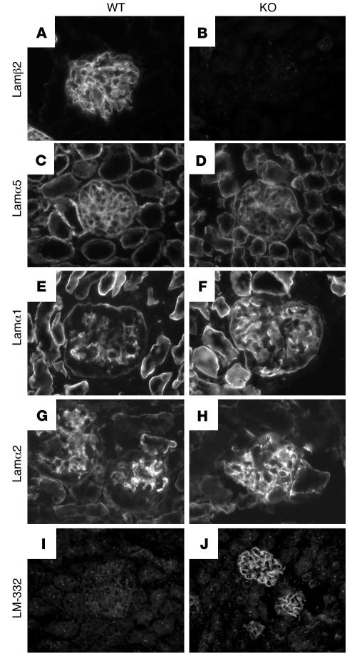
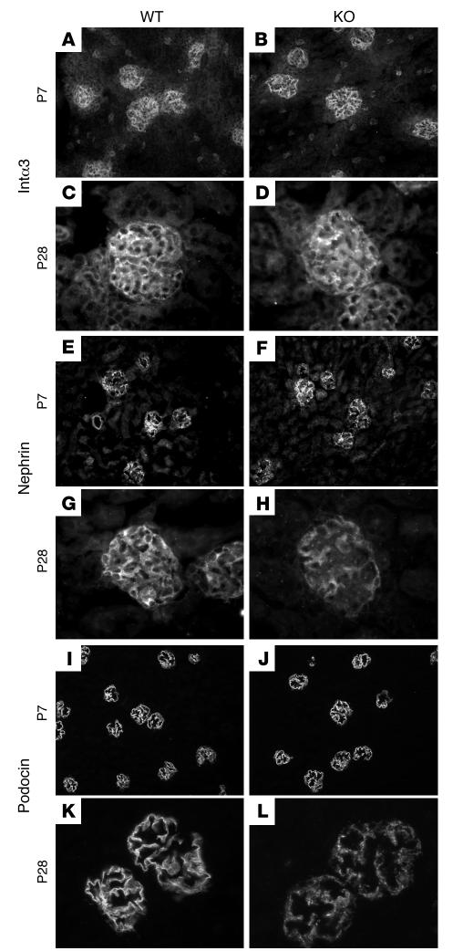
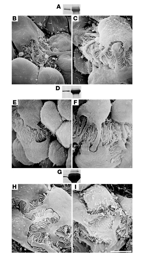
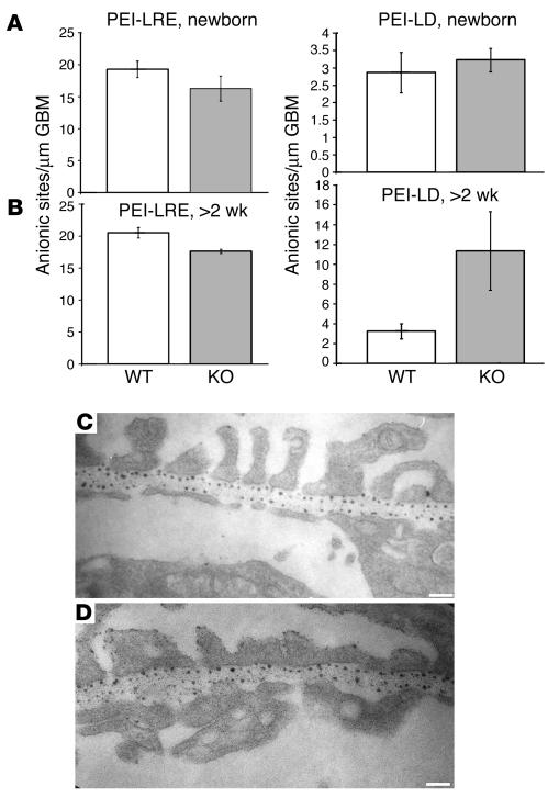
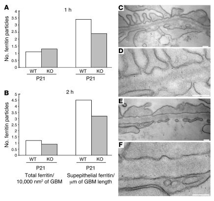
Primary defects in either podocytes or the glomerular basement membrane (GBM) cause proteinuria, a fact that complicates defining the barrier to albumin. Laminin beta2 (LAMB2) is a GBM component required for proper functioning of the glomerular filtration barrier. To investigate the GBM's role in glomerular filtration, we characterized GBM and overlying podocyte architecture in relation to development and progression of proteinuria in Lamb2-/- mice, which model Pierson syndrome, a rare congenital nephrotic syndrome. We found ectopic deposition of several laminins and mislocalization of anionic sites in the GBM, which together suggest that the Lamb2-/- GBM is severely disorganized, although it is ultrastructurally intact. Importantly, albuminuria was detectable shortly after birth and preceded podocyte foot process effacement and loss of slit diaphragms by at least 7 days. Expression and localization of slit diaphragm and foot process-associated proteins appeared normal at early stages. GBM permeability to the electron-dense tracer ferritin was dramatically elevated in Lamb2-/- mice, even before widespread foot process effacement. Increased ferritin permeability was not observed in nephrotic CD2-associated protein-null (Cd2ap-/-) mice, which have a primary podocyte defect. Together these data show that the GBM serves as a barrier to protein in vivo and that the glomerular slit diaphragm alone is not sufficient to prevent the passage of albumin into the urinary space.


Figures

Figure 1
Figure 1. Immunofluorescence detection of glomerular laminins.
Frozen sections of normal (left) and Lamb2–/–(right) kidneys were stained for laminin β2 (Lamβ2; A and B), α5 (C and D), α1 (E and F), α2 (G and H), and α3β3γ2 (LM-332; I and J). Normal GBM contains laminin β2 and α5 (and γ1; data not shown); laminin α5 was decreased in Lamb2–/–GBM. Normal glomeruli exhibit variable mesangial staining for laminin α1 and α2 but not LM-332; however, all were detected ectopically in Lamb2–/– GBM. Original magnification, ×600.
Figure 2
Figure 2. Immunofluorescence detection of podocyte proteins.
Frozen sections of normal (left) and Lamb2–/– (right) kidneys at P7 and P28 were stained for integrin α3 (Intα3; AD), nephrin (EH), and podocin (IL). There were no detectable changes in intensity or distribution of these proteins in young mice. However, with age and disease progression, there was a reduction in intensity; all 3 seemed to lose their normal linear distribution and showed a more diffuse granular pattern. This likely reflects the retraction of FPs and reduced SD density. Magnification, P7, ×200; P28, ×600.
Figure 3
Figure 3. Relationship between proteinuria and FP effacement inLamb2–/– glomeruli.
(A, D, and G) Equal volumes of urine from normal (left lanes) and Lamb2–/– (right lanes) littermates at P2, P5, and P10 (respectively) were analyzed by SDS-PAGE. Corresponding kidneys from normal (B, E, and H) and Lamb2–/– (C, F, and I) mice were analyzed by SEM. At P2 (B and C) and P5 (E and F), there were no signs of FP effacement, whereas areas of effacement were evident at P10 (H and I). Scale bar in I: 3 μm.
Figure 4
Figure 4. GBM anionic charge distribution.
Anionic sites were localized in normal and Lamb2–/– glomeruli (as indicated) at birth (A) and after 2 weeks of age (BD) using polyethylenimine. Anionic sites were counted and expressed as the number of sites/micrometer of GBM length in either the lamina densa (LD; middle of the GBM) or the lamina rara externa (LRE; podocyte aspect). There was a slight but consistent reduction in the total number of LRE anionic sites in the Lamb2–/– GBM, but an increase in the number of LD anionic sites at later ages, consistent with GBM disorganization. Data shown in A and B are mean ± SD. Scale bars: 125 nm.
Figure 5
Figure 5. Increased GBM permeability to ferritin inLamb2–/– mice.
Ferritin was detected in the GBM either 1 (A) or 2 (B) hour after a single intravenous injection into control/mutant littermate pairs, at the indicated ages. Ferritin particles were counted in the total surface area of the GBM (expressed as number of ferritin particles/10,000 nm2) or only at the subepithelial (podocyte) aspect (expressed as number of ferritin particles/μm of GBM length). There was an increase in total and in subepithelial ferritin at 1 hour in all Lamb2–/– mice compared with control or non-nephrotic, rescued Lamb2–/– mice carrying the NEPH-B2 transgene (Res). The increase was more remarkable after 2 hours in the older mice. (CF) Representative electron micrographs showing ferritin particles in the GBM of normal (C and D) and Lamb2–/– (E and F) mice at P11. Note the increased ferritin in the mutant GBM despite the normal FP architecture. D and F are higher magnifications of C and E, respectively. Scale bars: 125 nm.
Figure 6
Figure 6. Normal GBM permeability to ferritin inCd2ap–/– mice.
Ferritin injections were performed in Cd2ap–/– and control littermate mice. (A and B) Total numbers of ferritin particles in the GBM at 1 and 2 hours after injection were similar in nephrotic Cd2ap–/– mice and their normal littermates at P21. However, subepithelial ferritin particle numbers were lower in the mutant, despite the albuminuria. (CF) Representative electron micrographs showing ferritin particles in the GBM of P21 normal (C and D) and Cd2ap–/– (E and F) mice. D and F are higher magnifications of C and E, respectively. Scale bars: 125 nm.

Comment in

References

    1. Miner J.H., Yurchenco P.D. Laminin functions in tissue morphogenesis. Annu. Rev. Cell Dev. Biol. 2004;20:255–284. - PubMed
    1. Sasaki T., Fassler R., Hohenester E. Laminin: the crux of basement membrane assembly. J. Cell Biol. 2004;164:959–963. - PMC - PubMed
    1. Miner J.H. Renal basement membrane components. Kidney Int. 1999;56:2016–2024. - PubMed
    1. Abrahamson D.R. Origin of the glomerular basement membrane visualized after in vivo labeling of laminin in newborn rat kidneys. J. Cell Biol. 1985;100:1988–2000. - PMC - PubMed
    1. Aumailley M., et al. A simplified laminin nomenclature. Matrix Biol. 2005;24:326–332. - PubMed

Publication types