Comparative Study
. 2006 Oct 27;142(3):809-22.
doi: 10.1016/j.neuroscience.2006.06.045. Epub 2006 Aug 2.

Robust increase of cutaneous sensitivity, cytokine production and sympathetic sprouting in rats with localized inflammatory irritation of the spinal ganglia

Affiliations
Comparative Study

Robust increase of cutaneous sensitivity, cytokine production and sympathetic sprouting in rats with localized inflammatory irritation of the spinal ganglia

W-R Xie et al. Neuroscience. .

Abstract

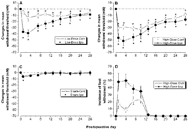
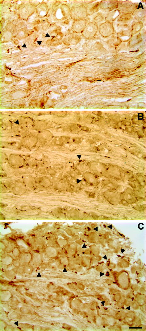
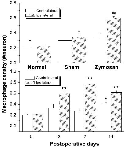
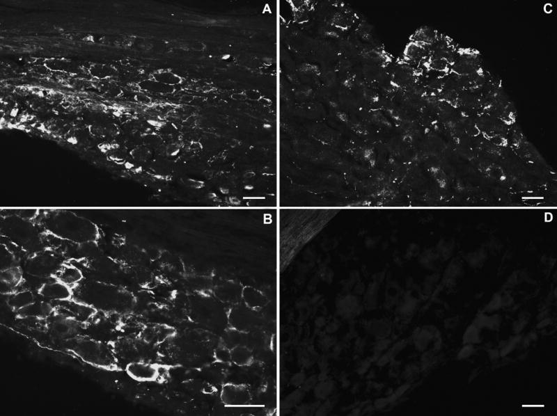
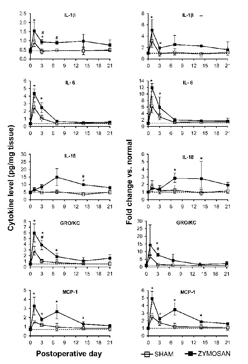
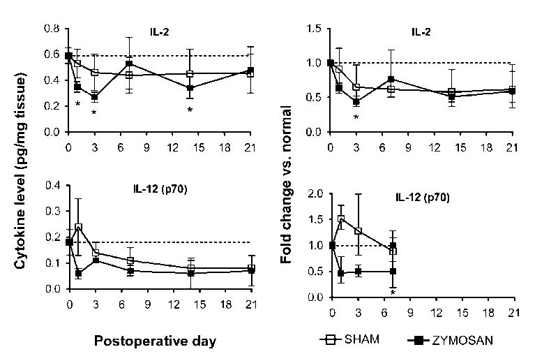
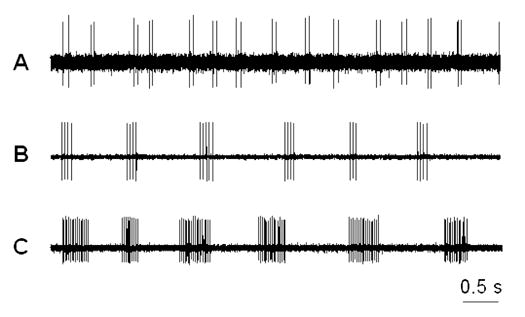
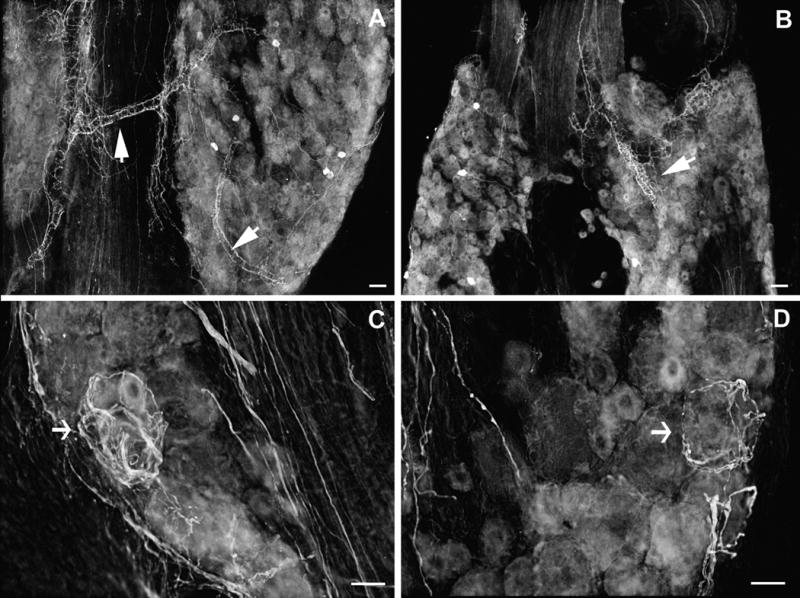
We investigated the role and mechanisms of inflammatory responses within the dorsal root ganglion (DRG) in the development of chemogenic pathological pain. DRG inflammation was induced by a single deposit of the immune activator zymosan in incomplete Freund's adjuvant in the epidural space near the L5 DRG via a small hole drilled through the transverse process. After a single zymosan injection, rats developed bilateral mechanical hyperalgesia and allodynia which began by day 1 after surgery, peaked at days 3-7, and lasted up to 28 days. The number of macrophages in ipsilateral and contralateral DRGs increased significantly, lasting over 14 days. Robust glial activation was observed in inflamed ganglia. Cytokine profile analysis using a multiplexing protein array system showed that, in normal DRG, all but interleukin (IL)-5, IL-10 and granulocyte-macrophage colony stimulating factor (GM-CSF) were detectable with concentrations of up to 180 pg/mg protein. Local inflammatory irritation selectively increased IL-1beta, IL-6, IL-18, monocyte chemoattractant protein-1 (MCP-1), and growth-related oncogene (GRO/KC) up to 17-fold, and decreased IL-2 and IL-12 (p70) up to threefold. Inflaming the DRG also remarkably increased the incidence of spontaneous activity of A- and C-fibers recorded in the dorsal root. Many of the spontaneously active A-fibers exhibited a short-bursting discharge pattern. Changes in cytokines and spontaneous activity correlated with the time course of pain behaviors, especially light stroke-evoked tactile allodynia. Finally, local inflammation induced extensive sprouting of sympathetic fibers, extending from vascular processes within the inflamed DRG. These results demonstrate the feasibility of inducing chronic localized inflammatory responses in the DRG in the absence of traumatic nerve damage, and highlight the possible contribution of several inflammatory cytokines/chemokines to the generation of spontaneous activity and development and persistence of chemogenic pathologic pain.
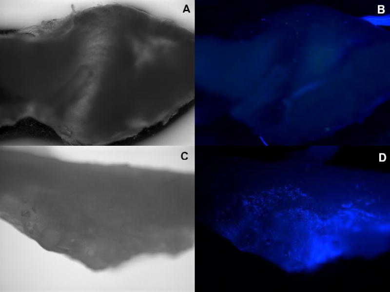

Figures

Figure 1
Figure 1
Inflaming the L5 DRG with zymosan/IFA induced prolonged bilateral mechanical hyperalgesia and allodynia. A, B. Changes in the withdrawal threshold to mechanical indentation of the hind paws in rats receiving local deposition of zymosan of 1 μg (low dose; A) or 10 μg (high dose; B) in IFA. Mean withdrawal thresholds to mechanical indentation are plotted as a function of postoperative time. Each data point is the mean threshold changes obtained on each day of testing. “Day 0” represents the withdrawal threshold changes averaged from 3 preoperative testing sessions (one session every 3 days). One-way RM ANOVA: Low-dose group, significant decreases from preoperative level (*p<0.05) beginning on the first postoperative day in the ipsilateral foot and on the third postoperative day in the contralateral side. High-dose group, significant decreases (*p<0.05) beginning on the first postoperative day in both the ipsilateral and contralateral foot. C. Control rats receiving local injection of buffered saline. D: Incidence of foot withdrawal response to light stroke of the ventral surface using a piece of cotton swab.
Figure 2
Figure 2
Light microscopic images of macrophage responses in zymosan-treated (10 μg) lumbar ganglia. Immunohistochemistry with ED-2 antibody on frozen sections (10 μm thick) of normal and zymosan-treated DRGs on POD 3. A: Example of a section from normal DRG with a few macrophages (arrow); B: representative example of an image from sham DRG on POD 3. C. DRG with immunological activation by injection (10 μg in IFA) of zymosan 3 days prior to immunostaining. Scale bar=50 μm.
Figure 3
Figure 3
Zymosan treatment (10 μg in IFA) induces prolonged macrophage response in the ipsilateral and, to a lesser degree, the contralateral DRGs. Top: Comparison of macrophage densities in the L5 DRGs subjected to various treatments. Immunohistochemistry with ED-2 antibody was performed in normal, sham, and Zymosan/IFA-treated DRG sections on postoperative day 3. Each data point is the mean density obtained from 8–10 sections of 3 ganglia from 3 rats. *P<0.05, Student’s t-test compared to normal. ##P<0.01, Student’s t-test compared to sham group. Bottom: Time course of macrophage response in the DRGs after zymosan/IFA deposit. *P<0.05, Student’s t-test compared to day “0”; **P<0.01, Student’s t-test compared to day “0”.
Figure 4
Figure 4
Light microscopic images of satellite glial activation (GFAP-positive) in zymosan/IFA-treated lumbar ganglia. Immunohistochemistry with anti- GFAP antibody was performed on frozen sections (10 μm thick) of normal and zymosan/IFA-treated DRGs on POD 3. A, B: L4 DRG ipsilateral to zymosan/IFA injection; C: L4 DRG contralateral to zymosan/IFA injection; D: normal, untreated DRG. Scale bar = 50 μm.
Figure 5
Figure 5
Selective up-regulation of cytokines in the inflamed DRGs. Time course of levels of the indicated cytokines is shown on the left; the normal values in unoperated rats are indicated by the dotted lines. Fractional changes (normal rat values used as the basis) are shown on the right. Zymosan dose was 10 μg.
Figure 6
Figure 6
Selective down-regulation of cytokines in the inflamed DRGs. Time course of levels of the indicated cytokines is shown on the left; the normal values in unoperated rats are indicated by the dotted lines. Fractional changes (normal rat values used as the basis) are shown on the right. Zymosan dose was 10 μg.
Figure 7
Figure 7
Three typical patterns of bursting discharge recorded from dorsal root fibers (Aβ) of the DRGs inflamed with Zymosan (10 μg). A: doublet discharges; B: short-bursting regular discharges; C: long-bursting discharges. The majority of spontaneously active neurons exhibited a short-bursting pattern.
Figure 8
Figure 8
Light microscopic photo images of the sprouted sympathetic fibers in the lumbar ganglia following localized inflammatory irritation with 10 μg Zymosan in IFA on postoperative day 7. A, B: TH-ir sympathetic fibers sprout from the vascular processes. C, D: sprouted fibers form basket structures around large DRG neurons. Images are inverted from the originals. Scale bar =50 μm.
None
Top (A, B): Contralateral L5 DRG Bottom (C, D): Ipsilateral L5 DRG Scale: 4x Note that the ipsilateral L5 DRG is covered by the fluospheres (bright spots).

References

    1. Barron KD, Marciano FF, Amundson R, Mankes R. Perineuronal glial responses after axotomy of central and peripheral axons. A comparison. Brain Res. 1990;523:219–229. - PubMed
    1. Beuche W, Friede RL. The role of non-resident cells in Wallerian degeneration. Journal of Neurocytology. 1984;13:767–796. - PubMed
    1. Briggs CA, Chandraraj S. Variations in the lumbosacral ligament and associated changes in the lumbosacral region resulting in compression of the fifth dorsal root ganglion and spinal nerve. Clin Anat. 1995;8:339–346. - PubMed
    1. Brown MC, Perry VH, Lunn ER, Gordon S, Heumann R. Macrophage dependence of peripheral sensory nerve regeneration: possible involvement of nerve growth factor. Neuron. 1991;6:359–370. - PubMed
    1. Burchiel KJ. Spontaneous impulse generation in normal and denervated dorsal root ganglia: sensitivity to alpha-adrenergic stimulation and hypoxia. Exp Neurol. 1984;85:257–272. - PubMed

Publication types

MeSH terms