. 2006 Nov 24;281(47):35686-98.
doi: 10.1074/jbc.M603357200. Epub 2006 Aug 3.

Nitrated fatty acids: Endogenous anti-inflammatory signaling mediators

Affiliations

Nitrated fatty acids: Endogenous anti-inflammatory signaling mediators

Taixing Cui et al. J Biol Chem. .

Abstract

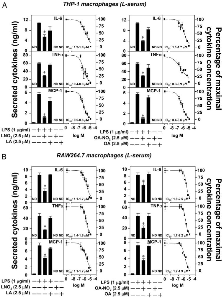
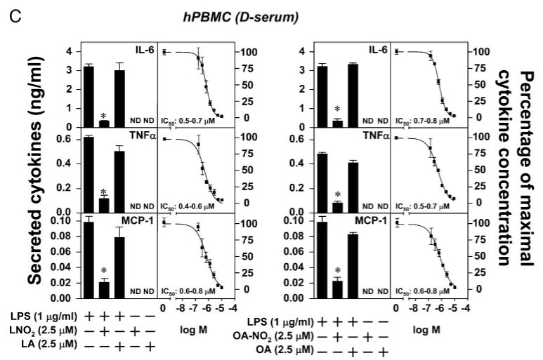
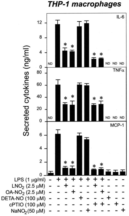
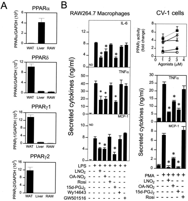
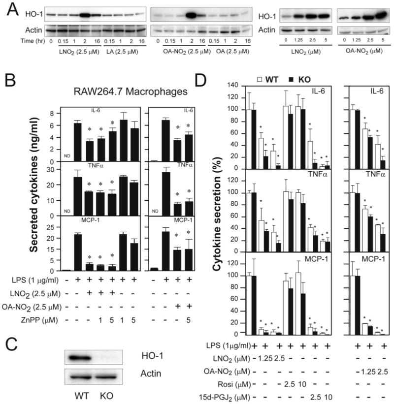
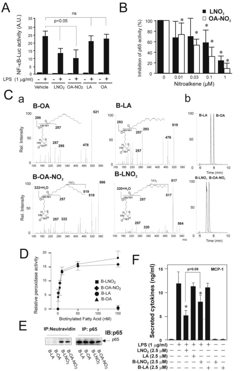
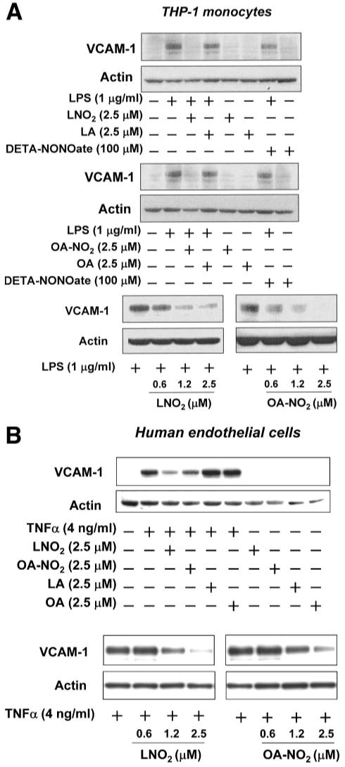
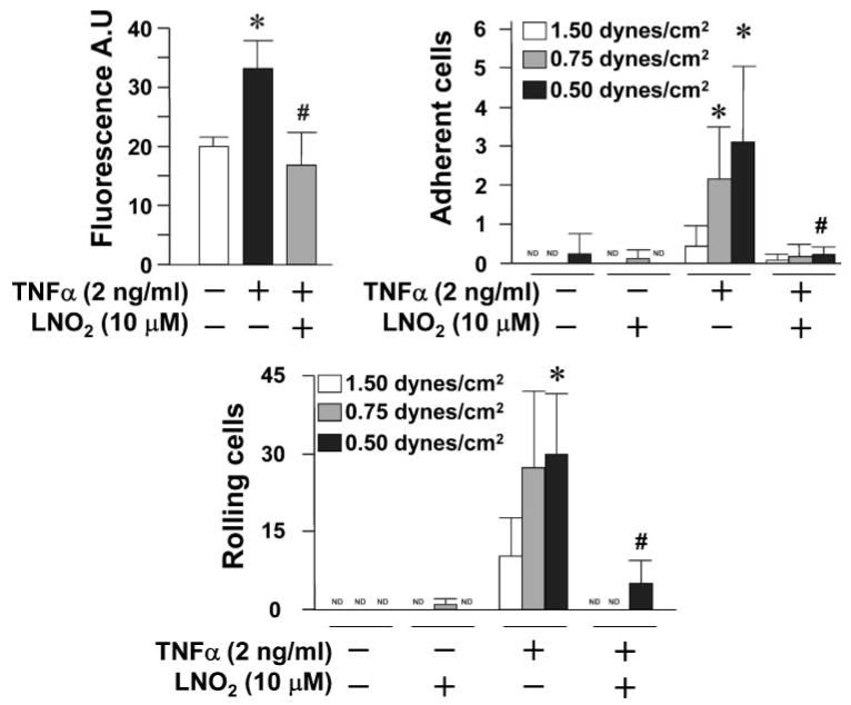
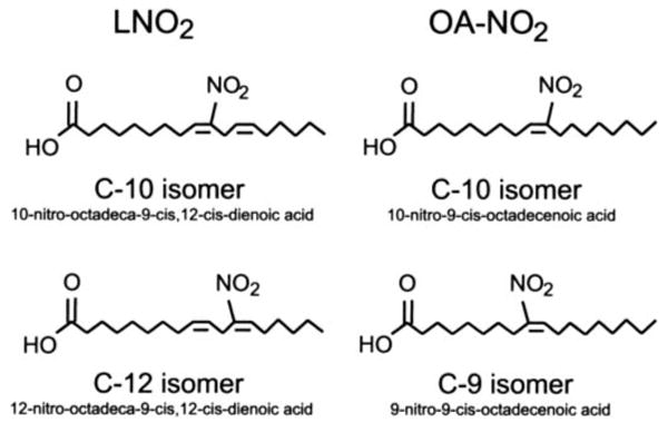
Nitroalkene derivatives of linoleic acid (LNO2) and oleic acid (OA-NO2) are present; however, their biological functions remain to be fully defined. Herein, we report that LNO2 and OA-NO2 inhibit lipopolysaccharide-induced secretion of proinflammatory cytokines in macrophages independent of nitric oxide formation, peroxisome proliferator-activated receptor-gamma activation, or induction of heme oxygenase-1 expression. The electrophilic nature of fatty acid nitroalkene derivatives resulted in alkylation of recombinant NF-kappaB p65 protein in vitro and a similar reaction with p65 in intact macrophages. The nitroalkylation of p65 by fatty acid nitroalkene derivatives inhibited DNA binding activity and repressed NF-kappaB-dependent target gene expression. Moreover, nitroalkenes inhibited endothelial tumor necrosis factor-alpha-induced vascular cell adhesion molecule 1 expression and monocyte rolling and adhesion. These observations indicate that nitroalkenes such as LNO2 and OA-NO2, derived from reactions of unsaturated fatty acids and oxides of nitrogen, are a class of endogenous anti-inflammatory mediators.


Figures

FIGURE 1
FIGURE 1. Nitrated fatty acids inhibit LPS-induced inflammatory cytokine secretion in macrophages
AC, LNO2 and OA-NO2 inhibited LPS-induced inflammatory cytokine secretion in THP-1 and RAW264.7 macrophages and human peripheral blood mononuclear cells (hPBMC) in either lipid-rich or delipidated serum. Cells were stimulated as indicated overnight in fresh culture medium with 1% lipid-rich normal serum (L-serum) (A and B) or with 1% delipidated serum (D-serum). The secretion of proinflammatory cytokines was assessed by ELISA. Values are expressed as mean ± S.D. (n = 6). *, p < 0.05 versus LPS alone. ND, nondetectable.
FIGURE 2
FIGURE 2. The inhibitory effect of LNO2 and OA-NO2 on LPS-induced proinflammatory cytokine secretion is NO-independent
Cells were stimulated overnight as indicated in fresh culture medium with 1% lipid-rich normal serum. DETA-NO represents DETA-NONOate. The secretion of proinflammatory cytokines was assessed by ELISA. Values are expressed as mean ± S.D. (n = 6). *, p < 0.05 versus LPS alone. ND, nondetectable.
FIGURE 3
FIGURE 3. Nitrated fatty acids inhibit secretion of proinflammatory cytokines in macrophages via PPARγ-independent pathways
A, relative expression levels of PPARs in RAW264.7 cells. Expression of PPARs was analyzed by real time PCR. Expression of PPARs in WAT and liver was used as positive control. B, comparison of PPAR ligands and nitrated lipids on the induced secretion of proinflammatory cytokines in RAW264.7 cells. Inset, the activity of PPARγ induced by various PPARγ ligands was measured using a CV-1 reporter assay (■, OA-NO2; ◆, LNO2; ▲, rosiglitazone (Rosi); ●, 15d-PGJ2; ○, vehicle). Proinflammatory cytokine production was measured by ELISA. The concentrations used in B were: LPS, 1 μg/ml; PMA, 0.1 μm; OA-NO2, 2.5 μm; LNO2, 2.5 μm; Rosiglitazone, 3 μm; 15d-PGJ2, 3 μm; GW501516, 0.5 μm; Wy14643, 100 μm. Values are expressed as mean ± S.D. (n = 6). *, p < 0.05 versus LPS alone. WAT, white adipose tissue of C57BL/6J mice; Liver, liver of C57BL/6J mice.
FIGURE 4
FIGURE 4. HO-1 is not essential for nitrated fatty acid-mediated anti-inflammatory signaling
A, time course and dose responses of nitrated fatty acid-induced HO-1 expression in RAW264.7 cells. B, effect of HO-1 inhibitor on nitrated fatty acid-mediated anti-inflammatory effect. Values are expressed as mean ± S.D. (n = 4). *, p< 0.05 versus LPS alone. C, expression of HO-1 in bone marrow-derived macrophages of HO-1 wild-type (WT) and knock-out (KO) mice. D, the anti-inflammatory effects of LNO2 and OA-NO2 in macrophages differentiated from bone marrow cells of HO-1 WT and KO mice. Values are expressed as mean ± S.D. (n = 4). *, p < 0.05 versus LPS alone. HO-1 expression was examined by Western blot analysis, and proinflammatory cytokine production was measured by ELISA as described under “Experimental Procedures.”
FIGURE 5
FIGURE 5. Nitrated fatty acids inhibit NF-κB activation by nitroalkylation of p65
A, LNO2 and OA-NO2 inhibit LPS-induced NF-κB activation in RAW264.7 cells. Cells were pretreated with LNO2 (2.5 μm) and OA-NO2 (2.5 μm) overnight and then stimulated with LPS (1 μg/ml) for 6 h. NF-κB activity was assessed as described under “Experimental Procedures.” Values are expressed as mean ± S.D. (n = 4). The data were analyzed using analysis of variance with the Newman-Keuls test. B, LNO2 and OA-NO2 directly inhibited NF-κB activity in vitro. Purified p65 was incubated with LNO2, OA-NO2, LA, and OA at different concentrations (0.01–1 μm) and then subjected to p65 activity assay as described under “Experimental Procedures.” Native fatty acids LA and OA at 0.01–1 μm had no effect on the DNA binding activity of p65. Therefore, the activity of p65 treated with LA (1 μm) or OA(1 μm) was used as a control and set to 100%. Values are expressed as mean ± S.D. (n = 4). The data were analyzed using analysis of variance with the Newman-Keuls test. C, structural characterization and chromatographic properties of biotinylated OA, LA, OA-NO2, and LNO2. a, structural characterization of biotinylated products by direct infusion into a ESI MS (ion trap) in negative ion mode. Main fragments are identified. Biotinylated lipids were prepared and characterized as described under “Experimental Procedures.” b, the corresponding elution profiles and retention times for the different biotinylated native and nitrated fatty acids are shown. D, p65 nitroalkylation by biotinylated LNO2 and OA-NO2 (B-LNO2 and B-OA-NO2, respectively) but not by native biotinylated fatty acids. Alkylation of p65 by nitroalkenes was assessed as described under “Experimental Procedures.” E, LNO2 and OA-NO2 covalently bind to p65 in vivo. RAW264.7 cells were treated with 0.1 μm biotinylated fatty acids and their corresponding nitro-derivatives, as indicated, for ∼1.5 h. Cell lysates were then treated with anti-p65 antibody or NeutroAvidin Plus followed by immunoblotting with anti-p65 antibody. F, biotinylated LNO2 inhibits LPS-induced inflammatory cytokine secretion in RAW264.7 macrophages. Cells were stimulated as indicated overnight, and the secretion of proinflammatory cytokines was assessed by ELISA. Values are expressed as mean ± S.D. (n = 3). *, p < 0.05 versus LPS alone.
FIGURE 6
FIGURE 6. Nitrated fatty acids inhibit the LPS- and TNF-α induced expression of VCAM-1 in THP-1 monocytes and human endothelial cells, respectively
A, LNO2 and OA-NO2 inhibited LPS-induced expression of VCAM-1 in THP-1 monocytes. B, LNO2 and OA-NO2 inhibited TNFα-induced expression of VCAM-1 in human endothelial cells. VCAM-1 expression was examined by Western blot analysis as described under “Experimental Procedures.”
FIGURE 7
FIGURE 7. LNO2 inhibits THP-1 monocyte adhesion and rolling on human endothelial cells
THP-1 monocyte adhesion and rolling on HUVEC was analyzed as described under “Experimental Procedures.” All values are expressed as mean ± S.D. (n = 4) and were representative of five independently performed experiments. The data were analyzed using Student's paired t test. *, p < 0.05 versus TNFα (−); #, p < 0.05 versus TNFα (+). ND, nondetectable.
SCHEME 1
SCHEME 1. Structures of LNO2 and OA-NO2
Structures of LNO2 and OA-NO2 used in this investigation are identical to those detected in the healthy human circulation (10, 11)

References

    1. Baldus S, Eiserich JP, Brennan ML, Jackson RM, Alexander CB, Freeman BA. Free Radic Biol Med. 2002;33:1010. - PubMed
    1. Ischiropoulos H, Beckman JS. J Clin Invest. 2003;111:163–169. - PMC - PubMed
    1. Rubbo H, Radi R, Trujillo M, Telleri R, Kalyanaraman B, Barnes S, Kirk M, Freeman BA. J Biol Chem. 1994;269:26066–26075. - PubMed
    1. Rubbo H, Parthasarathy S, Barnes S, Kirk M, Kalyanaraman B, Freeman BA. Arch Biochem Biophys. 1995;324:15–25. - PubMed
    1. O'Donnell VB, Eiserich JP, Chumley PH, Jablonsky MJ, Krishna NR, Kirk M, Barnes S, Darley-Usmar VM, Freeman BA. Chem Res Toxicol. 1999;12:83–92. - PubMed

Publication types

LinkOut - more resources