Differences between the scaling of miniature IPSCs and EPSCs recorded in the dendrites of CA1 mouse pyramidal neurons
- PMID: 16887875
- PMCID: PMC1995648
- DOI: 10.1113/jphysiol.2006.115428
Differences between the scaling of miniature IPSCs and EPSCs recorded in the dendrites of CA1 mouse pyramidal neurons
Abstract
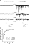
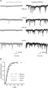
Anatomical studies have described inhibitory synaptic contacts on apical dendrites, and an abundant number of GABAergic synapses on the somata and proximal dendrites of CA1 pyramidal cells of the hippocampus. The number of inhibitory contacts decreases dramatically with distance from the soma, but the local electrophysiological characterization of these synapses at their site of origin in the dendrites is missing. We directly recorded dendritic GABA receptor-mediated inhibitory synaptic events in adult mouse hippocampal CA1 pyramidal neurons and compared them to excitatory synaptic currents recorded at the same sites. Miniature GABAergic events were evoked using localized application of a hyperosmotic solution to the apical dendrites in the vicinity of the dendritic whole-cell recording pipette. Glutamatergic synaptic events were blocked by kynurenic acid, leaving picrotoxin-sensitive IPSCs. We measured the amplitude and kinetic properties of mIPSCs at the soma and at three different dendritic locations. The amplitude of mIPSCs recorded at the various sites was similar along the somato-dendritic axis. The rise- and decay-times of local mIPSCs were also independent of the location of the synapses. The frequency of mIPSCs was 5 Hz at the soma, in contrast to < 0.5 Hz at dendritic sites, which could be increased to 10-20 Hz and 6-10 Hz, respectively, by our hyperosmotic stimulation protocol. Miniature glutamatergic events were evoked with the same protocol after blocking inhibitory synapses by bicucculine. The measured amplitudes increased along the somato-dendritic axis proportionally with their distance from the soma. The measured kinetic properties were independent of location. Consistent with the idea that IPSCs may have a restricted local effect in the dendrites, our data show a lack of distance-dependent scaling of miniature inhibitory synaptic events, in contrast to the scaling of excitatory events recorded at the same sites.
Figures


References
-
- Buhl EH, Halasy K, Somogyi P. Diverse sources of hippocampal unitary inhibitory postsynaptic potentials and the number of synaptic release sites. Nature. 1994;368:823–828. - PubMed
-
- Cossart R, Hirsch JC, Cannon RC, Dinoncourt C, Wheal HV, Ben-Ari Y, Esclapez M, Bernard C. Distribution of spontaneous currents along the somato-dendritic axis of rat hippocampal CA1 pyramidal neurons. Neuroscience. 2000;99:593–603. - PubMed
-
- Dinocourt C, Petanjek Z, Freund TF, Ben-Ari Y, Esclapez M. Loss of interneurons innervating pyramidal cell dendrites and axon initial segments in the CA1 region of the hippocampus following pilocarpine-induced seizures. J Comp Neurol. 2003;459:407–425. - PubMed
-
- Freund TF, Buzsaki G. Interneurons of the hippocampus. Hippocampus. 1996;6:347–470. - PubMed
Publication types
MeSH terms
Substances
LinkOut - more resources
Full Text Sources
Miscellaneous

