Comparative Study
. 2006 Aug 15;177(4):2452-62.
doi: 10.4049/jimmunol.177.4.2452.

Diverse immunoglobulin light chain organizations in fish retain potential to revise B cell receptor specificities

Affiliations
Comparative Study

Diverse immunoglobulin light chain organizations in fish retain potential to revise B cell receptor specificities

Ellen Hsu et al. J Immunol. .

Abstract

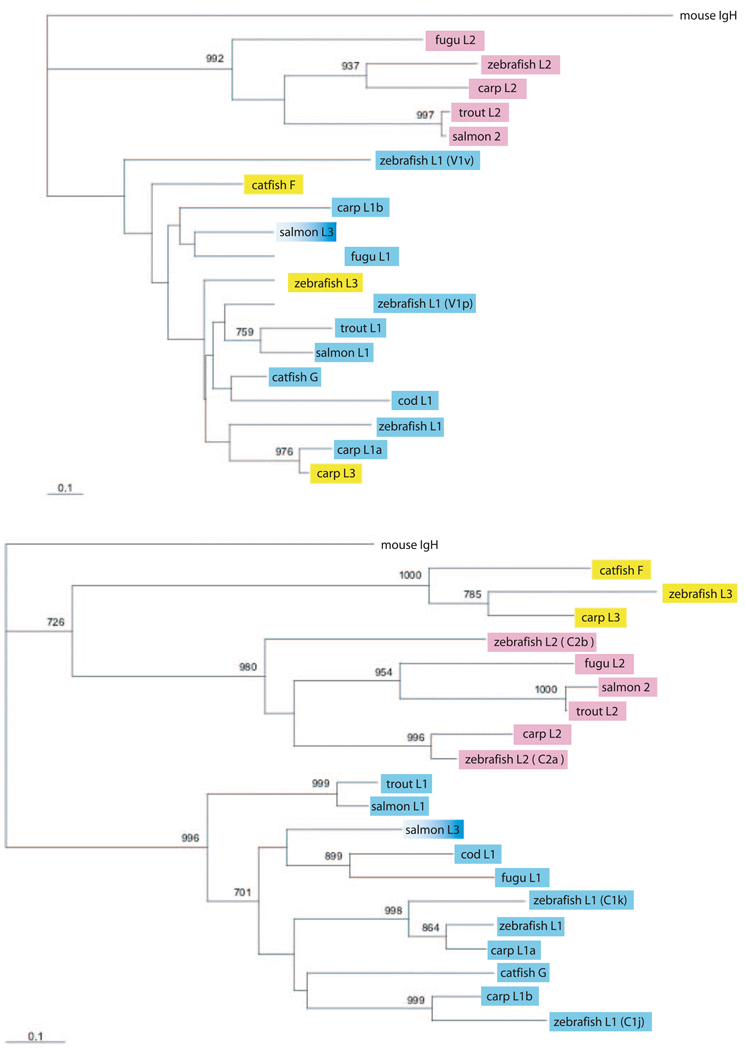
We have characterized the genomic organization of the three zebrafish L chain isotypes and found they all differed from those reported in other teleost fishes. Two of the zebrafish L chain isotypes are encoded by two loci, each carrying multiple V gene segments. To understand the derivation of these L chain genes and their organizations, we performed phylogenetic analyses and show that IgL organization can diverge considerably among closely related species. Except in zebrafish, the teleost fish IgL each contain only two to four recombinogenic components (one to three V, one J) and exist in multiple copies. BCR heterogeneity can be generated, but this arrangement apparently provides neither combinatorial diversification nor an opportunity for the secondary rearrangements that, in mammals, take place during receptor editing, a process crucial to the promotion of tolerance in developing lymphocytes. Examination of the zebrafish IgL recombination possibilities gave insight into how the suppression of self-reactivity by receptor editing might be managed, including in miniloci. We suggest that, despite the diverse IgL organizations in early and higher vertebrates, two elements essential to generating the Ab repertoire are retained: the numerous genes/loci for ligand-binding diversification and the potential for correcting unwanted specificities that arise.


Conflict of interest statement

Disclosures

The authors have no financial conflict of interest.

Figures

FIGURE 1
FIGURE 1
Organization of representative genes encoding zebrafish L chains. Five contigs from the zebrafish database were analyzed for the arrangement of L chain V (gray boxes) and J gene segments and C exons (black boxes). The transcriptional polarity is indicated by overhead arrows. Each gene is labeled, and an asterisk indicates presence of stop codon or discontinuous coding sequence. The sequences of functional genes and their positions are available in supplemental Fig. 1b. Type 1, Three contigs (NW_633979, NW_646408, NW_633913). NW_633979, The coding sequences of V1f/V1h 85% share identity and their leader introns 99% identity, suggesting that they are duplications. The complete sequence of V1j is not available from the current database. Most of these sequences closely resemble the four V genes on contig NW_64608: V1a is overall 85% identical with V1i, V1b 89% with V1k, and V1c is 97% identical with V1e. V1d is 92% identical with V1a. The end of the contig sequence is nearby; there probably exist additional upstream V genes, because a J gene segment (not shown) is present 10 kb upstream of V1a. NW_633913 contains a series of clusters extending over 516 kb. There are four functional C exons; the overall V sequences are well diverged from each other (31–100% identity) and from the nonlinked ones described above (31–67% identity). The V gene segments appear to be functional except for V1n, V1q, V1s, and V1u, which have stops; all RSS are intact. There is no J gene segment between V1p and C1g, nor is there a C exon between J1g and V1r. Type 2 (contig NW_644395), The functional V fall into two groups: V2b, V2e, V2f, V2g, V2h, V2k and V2a, V2c, V2d, V2i, V2j, V2l. Within a group, they are 89–95% at the nucleotide level in the V gene segment; between the two groups, they are 50–55% identical. Thus, these V genes are the least diverse among the three isotypes. We tried to ascertain how the four J gene segments are related by including the nearest upstream V gene in the sequence comparisons. A search using the sequence encompassing V2h-J2b (1144 bp) revealed that V2f-J2a share >90% identity over 920 bp, from the leader extending into the intervening DNA 450 bp beyond the RSS of the V gene. This suggests that they are relatively recent duplications. V2k is 90% identical with them, but homology drops off after the RSS. J2c also differs from the identical J2a/J2b/J2d, but the overall impression is that this type 2 locus arose from a series of tandem duplications. V2l-J2d are located 22 kb from C2b. All of the V gene segments appear to be functional except for V2b, which may have a nonfunctional RSS and V2i, which has an insertion in CDR3. Type 3 (contig NW_634729), The seven V3 gene segments overall share 46–94% nucleotide identity in the coding regions. The location of the V genes that are similar (V3a/V3h, 91%, and V3e/V3g, 95% identity) give little clue as to how the locus evolved; extensive changes have occurred that are difficult to reconstruct. V3a through V3f could have been generated by tandem duplication, but it is also possible that two clusters could have become merged, followed by deletion of a J and C formerly located after V3h. The V3b segment is in a different transcriptional orientation, but because the coding sequence is not outstanding, its position probably resulted from a meiotic inversion event. V3c is a pseudogene.
FIGURE 2
FIGURE 2
Recombination at the type 2 and type 3 loci. A, Deletion rearrangement at type 2 locus. B, Possible deletion rearrangement excising C2a exon. The excised region is indicated by brackets. C, Inversion rearrangement at type 3 (or type 1) locus. D, Inversion rearrangement to 3′ V gene segments in type 3 (or type 1) locus. Arrows show the sites of DNA breakage and inversion. The transcription polarity of the rearranged VJ, at the right, is indicated for A, C, and D. The RSS with 12-bp spacer is indicated as a black triangle, the RSS with 23-bp spacer is indicated as a white triangle.
FIGURE 3
FIGURE 3
Phylogenetic analyses of L chain V and C from various teleost fishes. Top, V domain. Bottom, C domain. All type 1/L1 sequences are indicated in blue, type 2/L2 in pink, and all type 3/L3 in yellow except salmon, which was named type 3, but which we designate as a type 1 (see Results). The following sequences were used in alignment. IGH, Mouse Mus musculus V MUSIGHVP, C MUSIGCD10. IGL, Channel catfish Ictalurus punctatus F (U25705, G L25533); zebrafish D. rerio L1(AF246185); L1(V1v) and L1(C1k) from NW_644842; L1(V1p) and L1(C1j) from NW_633913; L2 V (AF246183); L2(C2a) from NW_644395; L2(C2b) from NW_644395; L3 (AF246193); fugu pufferfish Takifugu rubripes (L1 AB126061, L2 M007644); rainbow trout Oncorhynchus mykiss (L1 X65260, L2 V OMU69988, L2 C AJ251648); carp Carpio cyprinus L1a V (AB073328); L1a C (AB015905); L1b V (AB073332); L1b C (AB035728); L2 V (AB091112); L2 C (AB091120); L3 V (AB073335); L3 C (AB035730); Atlantic salmon Salmo salar L1 (AF273012); L2 V (AF406963 and AF406964); L2 C (AF297518); L3 (AF406956); Atlantic cod Gadus morhua L1 (AF104898). Bootstrap values >70% are shown at nodes.
FIGURE 4
FIGURE 4
Taxonomic relationships among several teleost species. The taxonomic classifications of the teleost fishes listed in Table I are displayed according to superorder, order, and genus (48). The L chains that have been reported here and in the references in Table I are listed below the common name of the fish model (blue box). We propose that the type 3 (L3) is derived from type 1 (L1) in separate events in Ostariophysi and in Protacanthopterygii (pink ovals) (see Results). The carp L1A and L1B, considered by some investigators to be two isotypes (49), carry C regions that are highly related to zebrafish C1k and C1j, respectively (see Results and Fig. 3, bottom); we consider all these to be variants of type 1 (L1).
FIGURE 5
FIGURE 5
Receptor editing possibilities at fish IgL. A, Secondary rearrangements at the mouse κ locus. Recombination involving nested rearrangement of upstream V gene segment and downstream J gene segment can replace and delete unwanted VJ. Recombination between upstream V gene segment and RS element deletes C exon and inactivate locus, as does recombination between a heptamer (IRS) in the J-C intron and the RS element. After Moore et al. (35). B, Possible secondary rearrangement at a type 1 (or type 3) locus. Downstream V gene segments can rearrange with RSS in the signal joint to delete unwanted VJ. C, Possible secondary rearrangement among cod L chain clusters. Modeled after genomic clone CgL10, where enhancer activity was found 3′ of only the first cluster; it was proposed that one enhancer can regulate several clusters (29). With an unwanted VJ, rearrangements continue at the activated region. Another VJ recombination may occur, or the first VJ may be deleted, as shown, by rearrangement between the RSS in the signal joint and an inverted, downstream V gene segment. If the latter occurs first, the enhancer is deleted and the area may no longer be active. The RSS with 12-bp spacer is indicated as a black triangle, and the RSS with 23-bp spacer is indicated as a white triangle.

References

    1. Riblet R. Immunoglobulin heavy chain genes of mouse. In: Honjo T, Alt FW, Neuberger MS, editors. Molecular Biology of B Cells. Amsterdam: Elsevier Academic Press; 2004. pp. 19–26.
    1. Fugmann SD, Lee AI, Shockett PE, Villey IJ, Schatz DG. The RAG proteins and V(D)J recombination: complexes, ends, and transposition. Annu. Rev. Immunol. 2000;18:495–527. - PubMed
    1. Zachau HG. Immunoglobulin κ genes in human and mouse. In: Honjo T, Alt FW, Neuberger MS, editors. Molecular Biology of B Cells. Amsterdam: Elsevier Academic Press; 2004. pp. 27–36.
    1. Gay D, Saunders T, Camper S, Weigert M. Receptor editing: an approach by autoreactive B cells to escape tolerance. J. Exp. Med. 1993;177:999–1008. - PMC - PubMed
    1. Radic MZ, Erikson J, Litwin S, Weigert M. B lymphocytes may escape tolerance by revising their antigen receptors. J. Exp. Med. 1993;177:1165–1173. - PMC - PubMed

Publication types

MeSH terms

Substances