. 2006 Aug 15;103(33):12499-504.
doi: 10.1073/pnas.0605394103. Epub 2006 Aug 3.

Melanoma growth is reduced in fat-1 transgenic mice: impact of omega-6/omega-3 essential fatty acids

Affiliations

Melanoma growth is reduced in fat-1 transgenic mice: impact of omega-6/omega-3 essential fatty acids

Shuhua Xia et al. Proc Natl Acad Sci U S A. .

Abstract

An important nutritional question as to whether the ratio of omega-6 (n-6) to omega-3 (n-3) fatty acids plays a role in tumorigenesis remains to be clarified in well qualified experimental models. The recently engineered fat-1 mice, which can convert n-6 to n-3 fatty acids and have a balanced ratio of n-6 to n-3 fatty acids in their tissues and organs independent of diet, allow carefully controlled studies to be performed in the absence of potential confounding factors of diet and therefore are a useful model for elucidating the role of n-6/n-3 fatty acid ratio in tumorigenesis. We implanted mouse melanoma B16 cells into transgenic and WT littermates and examined the incidence of tumor formation and tumor growth rate. The results showed a dramatic reduction of melanoma formation and growth in fat-1 transgenic mice. The level of n-3 fatty acids and their metabolite prostaglandin E(3) (PGE(3)) were much higher (but the n-6/n-3 ratio is much lower) in the tumor and surrounding tissues of fat-1 mice than that of WT animals. The phosphatase and tensin homologue deleted on the chromosome 10 (PTEN) gene was significantly up-regulated in the fat-1 mice. In vitro experiments showed that addition of the n-3 fatty acid eicosapentaenoic acid or PGE(3) inhibited the growth of B16 cell line and increased the expression of PTEN, which could be partially attenuated by inhibition of PGE(3) production, suggesting that PGE(3) may act as an antitumor mediator. These data demonstrate an anticancer (antimelanoma) effect of n-3 fatty acids through, at least in part, activation of PTEN pathway mediated by PGE(3).


Conflict of interest statement

Conflict of interest statement: No conflicts declared.

Figures

Fig. 1.
Fig. 1.
Tumorigenicity of B16 melanoma cells in fat-1 transgenic and WT mice. (A) Different sizes of melanomas in WT and fat-1 transgenic (FAT-1) mice at two different time points. A number of 5 × 10⁶ viable cells in 50 μl of PBS were injected s.c. into each of 10 transgenic and 10 WT littermates (2-month-old female). On days 7 and 15 after cell implantation, animals were anesthetized briefly with isofluorane, and tumors were examined and photographed by using a digital camera. (B) Growth rates of melanomas in WT and transgenic mice. Tumor growth was monitored at the indicated time points by measuring the length, L, and width, w, of the tumor with a caliper and calculating tumor volume on the basis of the following formula: volume = (1/2)Lw2. The points are mean values ± SD of 10 tumors (n = 10) for the WT group or of 7 tumors (n = 7) for the fat-1 transgenic group (fat-1).
Fig. 2.
Fig. 2.
Identification of PGE2 (A; m/z 351) and PGE3 (B; m/z 349) by MS/MS in the tumor samples of fat-1 transgenic mice. LC-MS chromatograms showing the relative contents of PGE2 (C) and PGE3 (D) in melanoma and surrounding tissues of fat-1 transgenic and WT mice. Combined samples of three aliquots of tissues (tumor or stroma) from three different animals (fat-1 or WT) were extracted and analyzed for PGE2 and PGE3 by LC-MS/MS.
Fig. 3.
Fig. 3.
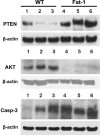
Western blotting of PTEN (Top), Akt (Middle), and caspase-3 (Bottom) in melanoma tumors from three WT (lanes 1–3) and three fat-1 transgenic (lanes 4–6) mice.
Fig. 4.
Fig. 4.
Effects of PGs (PGE2 and PGE3) on the growth of B16 melanoma cells and PTEN expression. (A) MTT assay of cell proliferation. B16 cells were treated with 1 μM PGE2 or 1 μM PGE3, and viable cells were determined at different time points by MTT assay. (B) Percentage of apoptotic cells after treatment with various concentrations of PGE3 for 72 h, determined by flow cytometry. (C) Western blot showing expression of PTEN. After treatment with 1 μM PGE2 or 1 μM PGE3 for 48 h, cells were harvested, and Western blot was performed to detect PTEN expression. β-Actin was used as control.
Fig. 5.
Fig. 5.
Effects of EPA, AA, and AA or EPA plus indomethacin on B16 cell viability (A) and cellular production of PGE2 and PGE3 (B). B16 cells were treated with 1% ethanol (as vehicle control), 50 μM AA, 50 μM AA plus 50 μM indomethacin, 50 μM EPA, and 50 μM EPA plus 50 μM indomethacin, respectively (in the presence of 10% FBS). After 48 h, cell viability was determined by staining cells with 1 μM calcein-AM and measuring the fluorescence intensity (n = 5); PGE2 and PGE3 were measured by LC-MS/MS (n = 4).

References

    1. Lands W. E. Am. Rev. Respir. Dis. 1987;136:200–204. - PubMed
    1. Samuelsson B., Dahlen S. E., Lindgren J. A., Rouzer C. A., Serhan C. N. Science. 1987;237:1171–1176. - PubMed
    1. James M. J., Gibson R. A., Cleland L. G. Am. J. Clin. Nutr. 2000;71:343S–348S. - PubMed
    1. Calder P. C. Ann. Nutr. Metab. 1997;41:203–234. - PubMed
    1. Rose D. P., Connolly J. M. Nutr. Cancer. 2000;37:119–127. - PubMed

Publication types

MeSH terms

LinkOut - more resources