. 2006 Aug 5:3:47.
doi: 10.1186/1742-4690-3-47.

The integrase interactor 1 (INI1) proteins facilitate Tat-mediated human immunodeficiency virus type 1 transcription

Affiliations

The integrase interactor 1 (INI1) proteins facilitate Tat-mediated human immunodeficiency virus type 1 transcription

Yasuo Ariumi et al. Retrovirology. .

Abstract

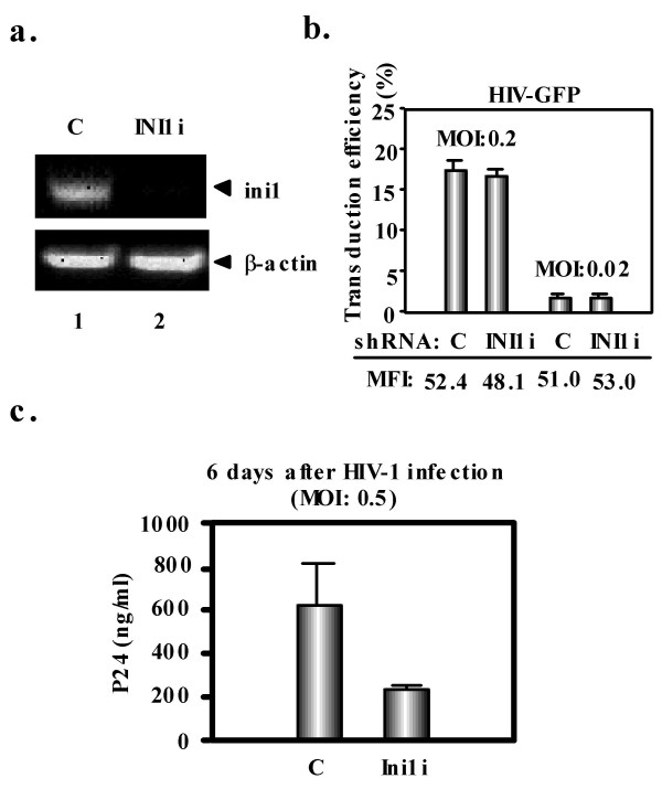
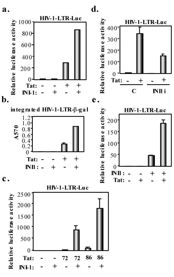
Integration of human immunodeficiency virus type 1 (HIV-1) into the host genome is catalyzed by the viral integrase (IN) and preferentially occurs within transcriptionally active genes. During the early phase of HIV-1 infection, the incoming viral preintegration complex (PIC) recruits the integrase interactor 1 (INI1)/hSNF5, a chromatin remodeling factor which directly binds to HIV-1 IN. The impact of this event on viral replication is so far unknown, although it has been hypothesized that it could tether the preintegration complex to transcriptionally active genes, thus contributing to the bias of HIV integration for these regions of the genome. Here, we demonstrate that while INI1 is dispensable for HIV-1 transduction, it can facilitate HIV-1 transcription by enhancing Tat function. INI1 bound to Tat and both the repeat (Rpt) 1 and Rpt 2 domains of INI1 were required for efficient activation of Tat-mediated transcription. These results suggest that the incoming PICs might recruit INI1 to facilitate proviral transcription.


Figures

Figure 1
Figure 1
INI1 is dispensable for HIV-1 transduction. a. Inhibition of ini1 expression by shRNA-producing lentiviral vector. Ethidium bromide-stained agarose gel analysis of RT-PCR amplification products of cytoplasmic RNA from INI1 knockdown P4.2 cells (INI1i) as well as in P4.2 cells transduced with a control (C) lentiviral vector. Cytoplasmic RNA was obtained using RNeasy kit (Qiagen) and RT-PCR was performed using SuperScript one-step RT-PCR with Platinum Taq kit (Invitrogen) with following primers: ini1, 5'-TCTGGAGGCGACTAGCCACTGTG-3' (forward primer) and 5'-GATCACAGCTGGGTCATGGTCATC-3' (reverse primer); beta-actin, 5'-TGACGGGGTCACCCACACTGTGCCCATCTA-3' (forward primer) and 5'-CTAGAAGCATTTGCGGTGGACGATGGAGGC-3' (reverse primer). b. Transduction efficiency in the same cells was determined by GFP fluorescence-activated cell sorter (FACS) analysis with a FACStrak apparatus (Becton Dickinson) 2 days after exposure to VSV-G-pseudotyped GFP-expressing HIV-1-derived lentiviral vector at the indicated MOI. Experiments were done in duplicate and columns represent the mean percentage of transduced cells, with mean fluorescence intensity (MFI) indicated below. c. Reduced HIV-1 replication in INI1 knockdown cells. INI1 knockdown P4.2 cells were infected with HIV-1 at a MOI of 0.5. HIV-1 replication was assayed by p24 ELISA in the culture supernatant 6 days later.
Figure 2
Figure 2
INI1 coactivates Tat-mediated HIV-1 transcription. a. P4.2 cells (2 × 10⁴ cells per well) were cotransfected with 100 ng of pHIV-1-LTR-Luc [25], 100 ng of pcDNA3-Tat101-FLAG [41], and/or 200 ng of pCGN-INI1 [7]. Luciferase assay was performed 24 h later. All transfections utilized equal total amounts of plasmid DNA owing to the addition of empty vector into the transfection mixture. Results were obtained through three independent transfections. Relative stimulation of luciferase activity (fold) is shown. b. 100 ng of pcDNA3-Tat101-FLAG, and/or 200 ng of pCGN-INI1 were cotransfected into P4.2 cells in triplicate. Beta-galactosidase activity was measured 24 h later. c. P4.2 cells were cotransfected with 100 ng of pHIV-1-LTR-Luc, 50 ng of pH2F Tat (Tat86) or pH2Tat (Tat72) [26], and/or 200 ng of pCGN-INI1 and performed luciferase assays 24 h later. d. 100 ng of pHIV-1-LTR-Luc, and/or 100 ng of pcDNA3-Tat101-FLAG were cotransfected into INI1 knockdown P4.2 cells (INI1i) or P4.2 cells transduced with a control (C) lentiviral vector and luciferase assays were performed 24 h later. e. INI1 deficient MON cells (2 × 10⁴ cells per well) were cotransfected with 100 ng of pHIV-1-LTR-Luc, 50 ng of pcDNA3-Tat101-FLAG, and/or 200 ng of pCGN-INI1 and luciferase assays performed 24 h later.
Figure 3
Figure 3
INI1 binds to Tat. 293T cells were cotransfected with 5 μg of pcDNA3-Tat101-FLAG [41] and/or 5 μg of pCGN-HA-INI1 [7]. Cells were lysed in buffer containing 50 mM Tris-HCl (pH 8.0), 150 mM NaCl, 4 mM EDTA, 0.1% NP-40, 10 mM NaF, 0.1 mM Na3VO4, 1 mM DTT and 1 mM PMSF. Lysates were pre-cleared with 30 μl of protein-G-sepharose (Amersham Biosciences). Pre-cleared supernatants were incubated with either 2 μg of anti-HA antibody (HA 11, Babco) or anti-FLAG antibody (M2, Sigma) at 4°C for 1 h. Following absorption of the precipitates on 30 μl of protein-G-sepharose resin for 1 h, the resin was washed four times with 700 μl lysis buffer. Bound proteins were eluted by boiling the resin for 5 min in 1× Laemmli sample buffer. The proteins were then subjected to SDS-PAGE, followed by immunoblotting analysis using either anti-HA or anti-FLAG antibodies.
Figure 4
Figure 4
Both Rpt1 and Rpt2 domains of INI1 are required for the efficient activation of Tat-mediated HIV-1 transcription. a. Schematic representation of HA-tagged full-length INI1 and its mutants. b. 293T cells (2 × 10⁴ cells per well) were cotransfected with 100 ng of pHIV-1-LTR-Luc [25], 50 ng of pcDNA3-Tat101-FLAG [41], and/or 200 ng of pCGN-INI1 or its mutant (20.2, 1.2, 3B, 27B) [7] in triplicate and luciferase assays were performed 24 h later. c. 293T cells were cotransfected with 5 μg of pcDNA3-Tat101-FLAG and/or 5 μg of pCGN-HA-INI1 or its mutant (20.2, 3B, 27B) and IP-western blotting was performed in the similar way as shown in Fig. 3. Asterisk denotes HA-INI1 or its mutant.

References

    1. Turlure F, Devroe E, Silver PA, Engelman A. Human cell proteins and human immunodeficiency virus DNA integration. Front Biosci. 2004;9:3187–3208. - PubMed
    1. Wang W, Côté J, Xue Y, Zhou S, Khavari PA, Biggar SR, Muchardt C, Kalpana GV, Goff SP, Yaniv M, Workman JL, Crabtree GR. Purification and biochemical heterogeneity of the mammalian SWI-SNF complex. EMBO J. 1996;15:5370–5382. - PMC - PubMed
    1. Kalpana GV, Marmon S, Wang W, Crabtree GR, Goff SP. Binding and stimulation of HIV-1 integrase by a human homolog of yeast transcription factor SNF5. Science. 1994;266:2002–2006. - PubMed
    1. Morozov A, Yung E, Kalpana GV. Structure-function analysis of integrase interactor 1/hSNF5L1 reveals differential properties of two repeat motifs present in the highly conserved region. Proc Natl Acad Sci USA. 1998;95:1120–1125. doi: 10.1073/pnas.95.3.1120. - DOI - PMC - PubMed
    1. Versteege I, Sévenet N, Lange J, Rousseau-Merck M-F, Ambros P, Handgretinger R, Aurias A, Delattre O. Truncating mutations of hSNF5/INI1 in aggressive paediatric cancer. Nature. 1998;394:203–206. doi: 10.1038/28212. - DOI - PubMed

Publication types

MeSH terms

LinkOut - more resources