

Advances in white matter imaging: a review of in vivo magnetic resonance methodologies and their applicability to the study of development and aging
- PMID: 16890990
- PMCID: PMC2895765
- DOI: 10.1016/j.neubiorev.2006.06.003
Advances in white matter imaging: a review of in vivo magnetic resonance methodologies and their applicability to the study of development and aging
Abstract
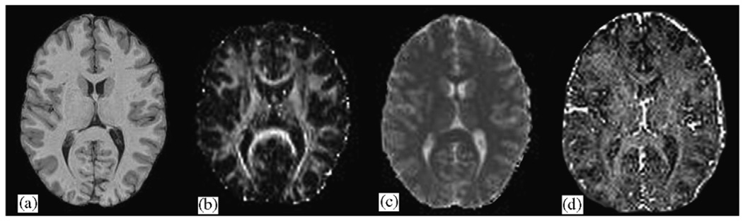
Several newer magnetic resonance imaging (MRI) techniques are increasingly being applied to the study of white matter development and pathology across the lifespan. These techniques go beyond traditional macrostructural volumetric methods and provide valuable information about underlying tissue integrity and organization at the microstructural and biochemical levels. We first provide an overview of white matter development and discuss the role of white matter and myelin in cognitive function. We also review available studies of development that have employed traditional volumetric measures. Then, we discuss the contributions of four newer imaging paradigms to our understanding of brain development and aging. These paradigms are Diffusion Tensor Imaging (DTI), Magnetization Transfer Imaging (MTI), T2-Relaxography, and Magnetic Resonance Spectroscopy (MRS). Studies examining brain development during childhood and adulthood as well as studies of the effects of aging are discussed.
Figures
Similar articles
-
A multimodal MRI approach to identify and characterize microstructural brain changes in neuropsychiatric systemic lupus erythematosus.Neuroimage Clin. 2015 May 16;8:337-44. doi: 10.1016/j.nicl.2015.05.002. eCollection 2015. Neuroimage Clin. 2015. PMID: 26106559 Free PMC article.
-
Arterial stiffness and white matter integrity in the elderly: A diffusion tensor and magnetization transfer imaging study.Neuroimage. 2019 Feb 1;186:577-585. doi: 10.1016/j.neuroimage.2018.11.015. Epub 2018 Nov 16. Neuroimage. 2019. PMID: 30448213
-
Texture analysis in brain T2 and diffusion MRI differentiates histology-verified grey and white matter pathology types in multiple sclerosis.J Neurosci Methods. 2022 Sep 1;379:109671. doi: 10.1016/j.jneumeth.2022.109671. Epub 2022 Jul 9. J Neurosci Methods. 2022. PMID: 35820450
-
The development of brain white matter microstructure.Neuroimage. 2018 Nov 15;182:207-218. doi: 10.1016/j.neuroimage.2017.12.097. Epub 2018 Jan 3. Neuroimage. 2018. PMID: 29305910 Free PMC article. Review.
-
In vivo quantification of white matter microstructure for use in aging: a focus on two emerging techniques.Am J Geriatr Psychiatry. 2014 Feb;22(2):111-21. doi: 10.1016/j.jagp.2013.08.001. Epub 2013 Sep 27. Am J Geriatr Psychiatry. 2014. PMID: 24080382 Free PMC article. Review.
Cited by
-
Structural modulation of brain development by oxygen: evidence on adolescents migrating from high altitude to sea level environment.PLoS One. 2013 Jul 9;8(7):e67803. doi: 10.1371/journal.pone.0067803. Print 2013. PLoS One. 2013. PMID: 23874449 Free PMC article.
-
Brain development in childhood.Open Neuroimag J. 2012;6:103-10. doi: 10.2174/1874440001206010103. Epub 2012 Nov 14. Open Neuroimag J. 2012. PMID: 23166579 Free PMC article.
-
Biomarkers of Aging and Relevant Evaluation Techniques: A Comprehensive Review.Aging Dis. 2024 May 7;15(3):977-1005. doi: 10.14336/AD.2023.00808-1. Aging Dis. 2024. PMID: 37611906 Free PMC article. Review.
-
Reproducibility of diffusion tensor imaging in normal subjects: an evaluation of different gradient sampling schemes and registration algorithm.Neuroradiology. 2014 Jun;56(6):497-510. doi: 10.1007/s00234-014-1342-2. Epub 2014 Mar 8. Neuroradiology. 2014. PMID: 24609528
-
Application of advanced neuroimaging modalities in pediatric traumatic brain injury.J Child Neurol. 2014 Dec;29(12):1704-17. doi: 10.1177/0883073814538504. Epub 2014 Jun 22. J Child Neurol. 2014. PMID: 24958007 Free PMC article. Review.
References
-
- Abernethy LJ, et al. Magnetic resonance imaging and T2 relaxometry of cerebral white matter and hippocampus in children born preterm. Pediatric Research. 2003;54(6):868–874. - PubMed
-
- Ashburner J, Friston KJ. Voxel-based morphometry—the methods. Neuroimage. 2000;11(6, Part 1):805–821. - PubMed
-
- Ashburner J, Friston KJ. Unified segmentation. Neuroimage. 2005;26:839–851. - PubMed
-
- Bagary MS, et al. Gray and white matter brain abnormalities in first-episode schizophrenia inferred from magnetization transfer imaging. Archives of General Psychiatry. 2003;60(8):779–788. - PubMed
Publication types
MeSH terms
Grants and funding
LinkOut - more resources
Full Text Sources
Medical
