. 2006 Aug;44(8):2921-32.
doi: 10.1128/JCM.00619-06.

Identification of Acinetobacter species and genotyping of Acinetobacter baumannii by multilocus PCR and mass spectrometry

Affiliations

Identification of Acinetobacter species and genotyping of Acinetobacter baumannii by multilocus PCR and mass spectrometry

Joseph A Ecker et al. J Clin Microbiol. 2006 Aug.

Abstract

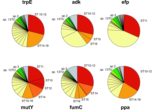
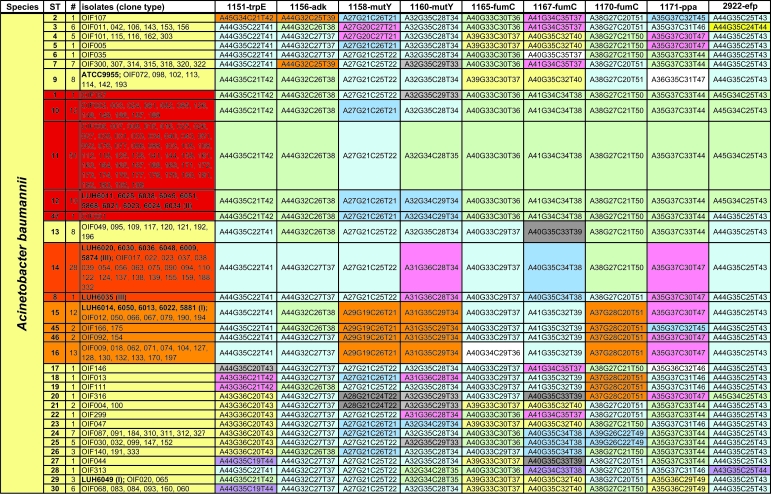
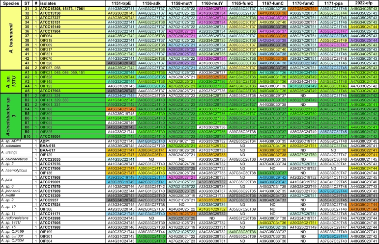
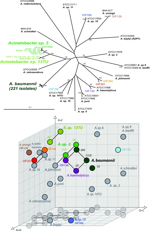
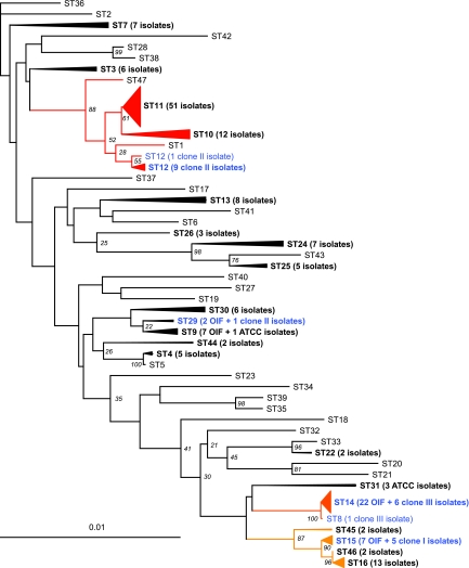
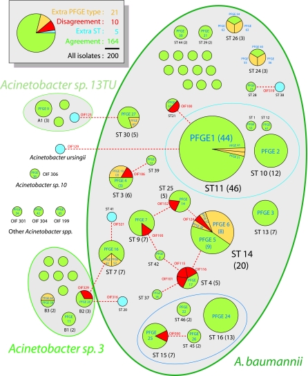
Members of the genus Acinetobacter are ubiquitous in soil and water and are an important cause of nosocomial infections. A rapid method is needed to genotype Acinetobacter isolates to determine epidemiology and clonality during infectious outbreaks. Multilocus PCR followed by electrospray ionization mass spectrometry (PCR/ESI-MS) is a method that uses the amplicon base compositions to genotype bacterial species. In order to identify regions of the Acinetobacter genome useful for this method, we sequenced regions of six housekeeping genes (trpE, adk, efp, mutY, fumC, and ppa) from 267 isolates of Acinetobacter. Isolates were collected from infected and colonized soldiers and civilians involved in an outbreak in the military health care system associated with the conflict in Iraq, from previously characterized outbreaks in European hospitals, and from culture collections. Most of the isolates from the Iraqi conflict were Acinetobacter baumannii (189 of 216 isolates). Among these, 111 isolates had genotypes identical or very similar to those associated with well-characterized A. baumannii isolates from European hospitals. Twenty-seven isolates from the conflict were found to have genotypes representing different Acinetobacter species, including 8 representatives of Acinetobacter genomospecies 13TU and 13 representatives of Acinetobacter genomospecies 3. Analysis by the PCR/ESI-MS method using nine primer pairs targeting the most information-rich regions of the trpE, adk, mutY, fumC, and ppa genes distinguished 47 of the 48 A. baumannii genotypes identified by sequencing and identified at the species level at least 18 Acinetobacter species. Results obtained with our genotyping method were essentially in agreement with those obtained by pulse-field gel electrophoresis analysis. The PCR/ESI-MS genotyping method required 4 h of analysis time to first answer with additional samples subsequently analyzed every 10 min. This rapid analysis allows tracking of transmission for the implementation of appropriate infection control measures on a time scale previously not achievable.


Figures

FIG. 1.
FIG. 1.
Distribution of the 267 Acinetobacter sequences into alleles for the six individual genes. Pie slices correspond to unique alleles. The area of the slice is determined by the number of isolates containing each allele. The color coding relates to sequence type groupings (Fig. 2), as follows: red, sequences belonging to A. baumannii group 11 (ST1, ST10, ST11, and ST12); dark orange, A. baumannii group 14 (ST8 and ST14); orange, A. baumannii group 15 (ST15, ST45, ST46, and ST16); yellow, all other A. baumannii types; medium green, Acinetobacter genomospecies 13TU; dark green, Acinetobacter genomospecies 3; white, other Acinetobacter species; gray, no sequence data available due to failed priming.
FIG.2.
FIG.2.
Base compositions of nine selected amplicons from Acinetobacter housekeeping genes. Within each column, base compositions that are common to multiple types are similarly colored. Base compositions printed on a white background are unique to a particular type. #, number of isolates; ND, base composition not determined.
FIG.2.
FIG.2.
Base compositions of nine selected amplicons from Acinetobacter housekeeping genes. Within each column, base compositions that are common to multiple types are similarly colored. Base compositions printed on a white background are unique to a particular type. #, number of isolates; ND, base composition not determined.
FIG. 3.
FIG. 3.
(Top) Phylogenetic relationships of Acinetobacter species as inferred from the analysis of 298 conserved positions of the efp gene. (Bottom) Distribution of the base compositions obtained for a primer pair encompassing 91 positions in efp. Since all amplicons have the same length, base compositions can be accurately mapped in a three-dimensional (G+C, A+C, A+G) space. The numbers of isolates that map to the same position are indicated in parentheses. Solid lines connect base compositions that are a single transition apart from one another. To facilitate depth perception, shadows are cast on the horizontal plane.
FIG. 4.
FIG. 4.
Phylogenetic relationships of Acinetobacter baumannii sequence types as inferred from the analysis of 1,679 conserved positions from the trpE, adk, efp, mutY, fumC, and ppa genes. Established clusters of types that differ in a single gene are indicated by red-orange shading.
FIG. 5.
FIG. 5.
Relationship between PCR/ESI-MS typing and PFGE genotyping. Both typing methods were used to analyze 200 isolates, and the isolates were distributed among pies in accordance with their sequence types (black labels). Within each pie, isolates were segregated according to PFGE types (blue labels). Green slices or pies represent agreement between typing methods. Yellow slices indicate isolates that are further discriminated by PFGE relative to the PCR/ESI-MS typing method. Blue pies represent isolates that are further discriminated by PCR/ESI-MS typing relative to PFGE. Red slices indicate disagreement between the PFGE and PCR/ESI-MS typing methods; red dashed lines connect the PCR/ESI-MS type with the PFGE type that was actually observed. Ellipses cluster together closely related types.

References

    1. Al-Khoja, M. S., and J. H. Darrell. 1979. The skin as the source of Acinetobacter and Moraxella species occurring in blood cultures. J. Clin. Pathol. 32:497-499. - PMC - PubMed
    1. Baltimore, R. S., R. L. Duncan, E. D. Shapiro, and S. C. Edberg. 1989. Epidemiology of pharyngeal colonization of infants with aerobic gram-negative rod bacteria. J. Clin. Microbiol. 27:91-95. - PMC - PubMed
    1. Bartual, S. G., H. Seifert, C. Hippler, M. A. Luzon, H. Wisplinghoff, and F. Rodriguez-Valera. 2005. Development of a multilocus sequence typing scheme for characterization of clinical isolates of Acinetobacter baumannii. J. Clin. Microbiol. 43:4382-4390. - PMC - PubMed
    1. Baumann, P. 1968. Isolation of Acinetobacter from soil and water. J. Bacteriol. 96:39-42. - PMC - PubMed
    1. Bergogne-Berezin, E. 2001. The increasing role of Acinetobacter species as nosocomial pathogens. Curr. Infect. Dis. Rep. 3:440-444. - PubMed

Publication types

MeSH terms

LinkOut - more resources