. 2006 Aug 7:3:48.
doi: 10.1186/1742-4690-3-48.

Effect of SWI/SNF chromatin remodeling complex on HIV-1 Tat activated transcription

Affiliations

Effect of SWI/SNF chromatin remodeling complex on HIV-1 Tat activated transcription

Emmanuel Agbottah et al. Retrovirology. .

Abstract

Background: Human immunodeficiency virus type 1 (HIV-1) is the etiologic agent of acquired immunodeficiency virus (AIDS). Following entry into the host cell, the viral RNA is reverse transcribed into DNA and subsequently integrated into the host genome as a chromatin template. The integrated proviral DNA, along with the specific chromatinized environment in which integration takes place allows for the coordinated regulation of viral transcription and replication. While the specific roles of and interplay between viral and host proteins have not been fully elucidated, numerous reports indicate that HIV-1 retains the ability for self-regulation via the pleiotropic effects of its viral proteins. Though viral transcription is fully dependent upon host cellular factors and the state of host activation, recent findings indicate a complex interplay between viral proteins and host transcription regulatory machineries including histone deacetylases (HDACs), histone acetyltransferases (HATs), cyclin dependent kinases (CDKs), and histone methyltransferases (HMTs).

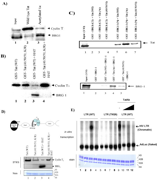
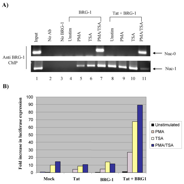
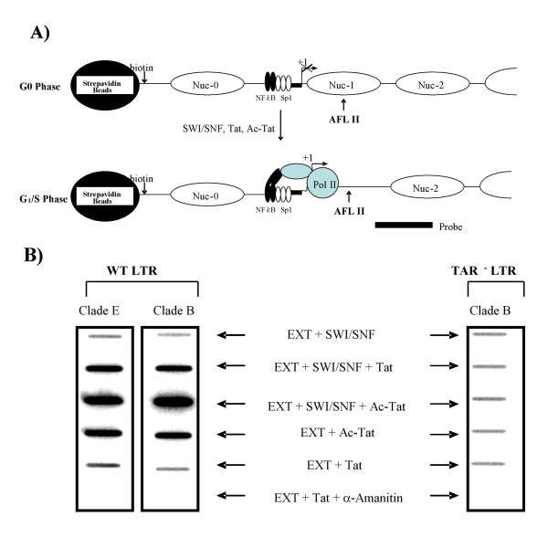
Results: Here, we describe the effect of Tat activated transcription at the G1/S border of the cell cycle and analyze the interaction of modified Tat with the chromatin remodeling complex, SWI/SNF. HIV-1 LTR DNA reconstituted into nucleosomes can be activated in vitro using various Tat expressing extracts. Optimally activated transcription was observed at the G1/S border of the cell cycle both in vitro and in vivo, where chromatin remodeling complex, SWI/SNF, was present on the immobilized LTR DNA. Using a number of in vitro binding as well as in vivo chromatin immunoprecipitation (ChIP) assays, we detected the presence of both BRG1 and acetylated Tat in the same complex. Finally, we demonstrate that activated transcription resulted in partial or complete removal of the nucleosome from the start site of the LTR as evidenced by a restriction enzyme accessibility assay.

Conclusion: We propose a model where unmodified Tat is involved in binding to the CBP/p300 and cdk9/cyclin T1 complexes facilitating transcription initiation. Acetylated Tat dissociates from the TAR RNA structure and recruits bromodomain-binding chromatin modifying complexes such as p/CAF and SWI/SNF to possibly facilitate transcription elongation.


Figures

Figure 1
Figure 1
HIV chromatin transcription at various stages of the cell cycle. To obtain synchronized extracts, both eTat and control cells were first treated with hydroxyurea, released and then treated with nocodazole. Extracts were made from 1 h (G0), 3 h (early G1), 6 h (G1/S), 15 h (late S), and 21 h (G2) post-nocodazole release and used in an in vitro transcription reaction. Released cells (1/10) were processed for FACS profile (data not shown). Two micrograms of chromatin DNA were used in each reaction. The bottom panels are gels of histones from immobilized DNA after transcription stained with Coomasie Blue. Lanes 1–5 and 11–15 are using eTat extract, and lanes 6–10 are using control pCEP4 extract. The left panel contains HIV-LTR-TAR wild-type and HIV-LTR-TAR mutant (TM26), and 200 ng of naked DNA (AdLuc, [55]).
Figure 2
Figure 2
HIV-1 Nef and Env expression in individual cells at various phases of the cell cycle. A) HIV-1 protein expression was monitored by flow cytometric analysis. OM10.1 cells were washed in Hanks' balanced salt solution, fixed in 70% ice-cold ethanol, and stained with mouse anti-human monoclonal antibodies to cyclin E, cyclin A, Nef, Env, or a non-specific immunoglobulin (DAKO) overnight. Cells in G1 and G2 were identified by DNA content and divided the intervening region into 10 to 12 equal increments to allow the calculation of mean protein-associated fluorescence and mean DNA content. The exact same analysis was performed on the appropriate aliquot stained with non-specific immunoglobulin. Protein fluorescence was determined by subtracting the mean from the nonspecific samples. Samples were washed in PBS-BSA and stained with a fluorescein isothiocyanate-labeled goat anti-mouse secondary antibody (1:30; DAKO) for 30 min in the dark. The bottom of panel A shows HIV-1 gene expression in G0, early G1, G1/S, S, and G2 phases of cell cycle by RT-PCR with primers for Env and Nef [58, 59]. B) Cell cycle analysis of cells after G0 release. Cells were synchronized at G0 by serum starvation and samples were removed from the medium at each time point, washed with PBS without Mg2+ or Ca2+, fixed with 70% ethanol, and stained with propidium iodide followed by cell-sorting analysis on a Coulter EPICS cell analyzer. The FACS profile for each time point is presented along with the percentage of cells in G1, S, and G2 (upper right side boxes of each histogram).
Figure 3
Figure 3
Identification of cellular proteins associated with the transcriptionally active nucleosomal HIV-1 promoter. A) Nucleosomes were assembled on biotin labeled HIV-1 LTR DNA and G1/S extracts were added to the reaction mixture. Following washes (TNE600 plus 1% NP-40), samples were eluted with excess biotin and centrifuged to pellet the beads. Supernatants were TCA precipitated and ran on a 4–20% gel and silver stained (MALDI compatible). Lane 1, Strepavidin beads; lane 2, purified core histones before nucleosomal assembly; lane 3, purified Tat; lanes 4–7 are LTR sequences (-110/+180) from four different clade B isolates assembled and incubated with G1/S extracts without any nucleotide addition; lane 8, transcriptionally active HXB2 promoter with nucleotide addition (ATP, CTP, GTP); lane 9, HXB2 promoter with purified Tat; lane 10 same as lane 8 with purified Tat; lane 11, the rainbow molecular marker (14–220 kDa). B) Control gel utilizing HXB2 wild-type, TATA-/TAR+, and TATA+/TAR- promoters with Tat and nucleotide (ATP, CTP, GTP) and AdML promoters. Unique proteins (.) bound to DNA were cut out, trypsin digested, and used for identification by mass spectrometry [74].
Figure 4
Figure 4
BRG1 binds to acetylated Tat. A) CEM G1/S cell extracts (2 mg) were mixed with 100 μg of biotin-Tat peptides (aa 42–54), incubated for 2 h, and washed. Bound proteins were separated on a 4–20% SDS/PAGE and Western blotted. Lane 1, input; lane 2, unacetylated Tat peptide; and lane 3, acetylated Tat peptide. B) GST-Tat (wild-type and mutant) was purified over a glutathione column and eluted. Acetylated GST-Tat was incubated with p300 and acetyl-CoA (62), washed, and incubated with cyclin T1/cdk9 (top) or SWI/SNF (bottom). Bound complexes were run on a 4–20% SDS/PAGE and Western blotted for the presence of cyclin T1 or BRG1. Lane 1, wild-type Tat; lane 2, Tat with a lysine to arginine change at residues 41, 50, and 51; lane 3, wild-type Tat and p300; lane 4, mutated Tat and p300. C) GST proteins and TNT lysates containing 35S-labeled Tat or BRG1 were incubated for 2 h at 4°C. Complexes were centrifuged; bound labeled proteins were denatured, subjected to SDS-PAGE, dried, and autoradiographed. D) Top: Schematic of the biotinylated HIV-1 LTR DNA used in the pull-down assay. Bottom: Immobilized chromatin HIV-1 LTR templates were incubated with CEM extract and wild-type or mutated Tat, then acetylated in the presence of GST-HAT. Samples were incubated with 100 ng SWI/SNF and all four cold nucleotides. Templates were washed and proteins were separated on a 4–20% SDS/PAGE for Western blot analysis. Bottom: remaining histones after transcription. E) CEM cells were treated with hydroxyurea and nocodazole, and samples were processed at 9 h post-release. The extracts were supplemented with purified SWI/SNF (all lanes), plus wild-type Tat (lanes 2 and 6), acetylated Tat (lanes 3 and 7), and Tat mutated at positions 50/51 (lanes 4 and 8). Anti-acetyl Tat 50/51 antibodies were added at time zero (lanes 10–12). Bottom: histone stain from immobilized DNA after transcription.
Figure 5
Figure 5
Acetylated Tat associates with a chromatinized HIV-1 promoter at or near nuc-1 but not nuc-1. A) Diagram of nucleosomes positioned on the integrated HIV-1 genome. The transcription start site is indicated as +1. Critical transcription factor binding sites (NF-κB, Sp1, and TBP) are indicated. Location of nuc-0 through nuc-4 are indicated above the diagram. B) ChIP analysis of the HIV-1 genome. The ChIP assay was performed as described [110, 111] with modifications (see Materials and Methods). HLM-1 cells (Tat-) [112] were transfected with the pCEP4/eTat vector [53] or no DNA and ChIP analysis was performed with anti- acetyl Tat 50/51 antibodies, anti-acetyl Tat 41 antibodies, or anti-acetyl H4 antibodies (positive control for acetylation of histones and association of nucleosomes with the HIV-1 promoter). The PCR primer pair location and expected sizes are indicated next to the expected amplified PCR products.
Figure 6
Figure 6
Suppression of BRG1 expression by RNAi inhibits HIV-1 replication. Complete sequence of BRG1 wild-type and mutant siRNA used in transfection. Oligos were designed and synthesized using the OligoEngine website [109]. HIV-1 infected cells (ACH2, OM10.1 and 8E5) were treated with TNF-α (10 ng/ml) for 2 h, washed, and subsequently electroporated (ACH2, [113]) or Amexa (OM10.1 and 8E5) treated with increasing amounts (1, 5, or 10 μg) of either wild-type (WT) or mutant (mut) BRG1 siRNA. Forty eight hours later, samples were collected and used for p24 gag ELISA. Western blot against BRG1 showed more than 90% decrease in protein levels in WT and not mutant BRG1 siRNA transfect cells [74].
Figure 7
Figure 7
BRG1 and Tat associate with chromatinized HIV-1 promoter in C33A cells at nuc-1. A) The BRG1/hBrm-deficient cervical carcinoma cell line C33A was used for these experiments and maintained under standard conditions in Dulbecco's modified Eagle's medium. A BRG1 (10 μg) or Tat (3 μg) expression vector or a control vector (10 μg, lane 3) was transfected into C33A cells that have an HIV-1 reporter construct stably integrated into their genome. After cells were allowed to express BRG1 for 36 h, cultures were stimulated with either PMA for 2 h (lane 5), TSA for 12 h (lane 6), or both for 2 h and 12 h, respectively (lane 7), before ChIP. A ChIP assay was performed with BRG1 antibodies, and PCR was used to detect the recovery of nuc-0 and nuc-1 DNA. The input DNA represents the total genomic DNA. Ab, antibody; Unstim, unstimulated. B) BRG1 and Tat were transiently expressed in C33A cells that had been stably transfected with an HIV-1-luciferase construct. Thirty-six hours after transfection, cells were stimulated with either PMA for 2 h, TSA for 12 h, or both for 12 h. Following stimulation, cells were lysed, and the luciferase activity was measured [114]. Data presented is from average of two individual experiments.
Figure 8
Figure 8
Effect of SWI/SNF, Tat, or acetylated Tat on restriction enzyme accessibility. A) A diagram of the transcription experiment. Immobilized templates (800 bp from the 5' U3 region and into the Gag region) were assembled into nucleosomes and SWI/SNF, Tat, or acetylated Tat were added to the reaction. In the absence of transcription, nuc-1 blocks restriction enzyme accessibility of Afl II. During transcription and remodeling of the nucleosome, the Afl II site becomes accessible, and the 3' end sequence is released. This fragment can be detected by southern slot blotting [74] using a probe (end labeled oligonucleotide) spanning the U5 region into Gag. B) In vitro transcription using CEM cells (G1/S extract). Similar to Figure 1, CEM cells were treated with hydroxyurea/nocodazole and samples were processed at 9 h post-release. The extracts (EXT, 200 μg) were supplemented with 200 ng of purified SWI/SNF, plus wild-type Tat (500 ng), or acetylated Tat (500 ng). α-amanitin at 0.1 μg/ml was used to detected RNAPII sensitivity. Clades B and E (a generous gift of J. Hiscott) and the TAR mutant LTR (TM26) were used as the DNA templates.

Similar articles

Cited by

References

    1. Fauci AS. Host factors in the pathogenesis of HIV disease. Antibiot Chemother. 1996;48:4–12. - PubMed
    1. Garza HHJ, Carr DJ. Interactions of human immunodeficiency virus type 1 transactivator of transcription protein with signal transduction pathways. Adv Neuroimmunol. 1995;5:321–325. doi: 10.1016/0960-5428(95)00014-S. - DOI - PubMed
    1. Coull JJ, Romerio F, Sun JM, Volker JL, Galvin KM, Davie JR, Shi Y, Hansen U, Margolis DM. The human factors YY1 and LSF repress the human immunodeficiency virus type 1 long terminal repeat via recruitment of histone deacetylase 1. J Virol. 2000;74:6790–6799. doi: 10.1128/JVI.74.15.6790-6799.2000. - DOI - PMC - PubMed
    1. Bohan CA, Kashanchi F, Ensoli B, Buonaguro L, Boris-Lawrie KA, Brady JN. Analysis of Tat transactivation of human immunodeficiency virus transcription in vitro. Gene Expr. 1992;2:391–407. - PMC - PubMed
    1. Feinberg MB, Baltimore D, Frankel AD. The role of Tat in the human immunodeficiency virus life cycle indicates a primary effect on transcriptional elongation. Proc Natl Acad Sci U S A. 1991;88:4045–4049. doi: 10.1073/pnas.88.9.4045. - DOI - PMC - PubMed

Publication types

MeSH terms

LinkOut - more resources