. 2006;8(5):R139.
doi: 10.1186/ar2029.

Activation of WNT and BMP signaling in adult human articular cartilage following mechanical injury

Affiliations

Activation of WNT and BMP signaling in adult human articular cartilage following mechanical injury

Francesco Dell'Accio et al. Arthritis Res Ther. 2006.

Abstract

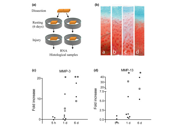
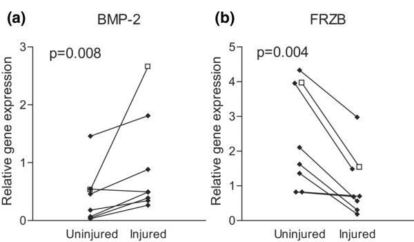
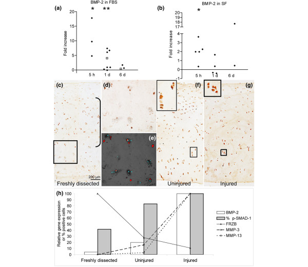
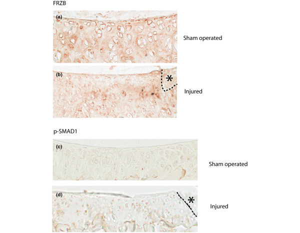
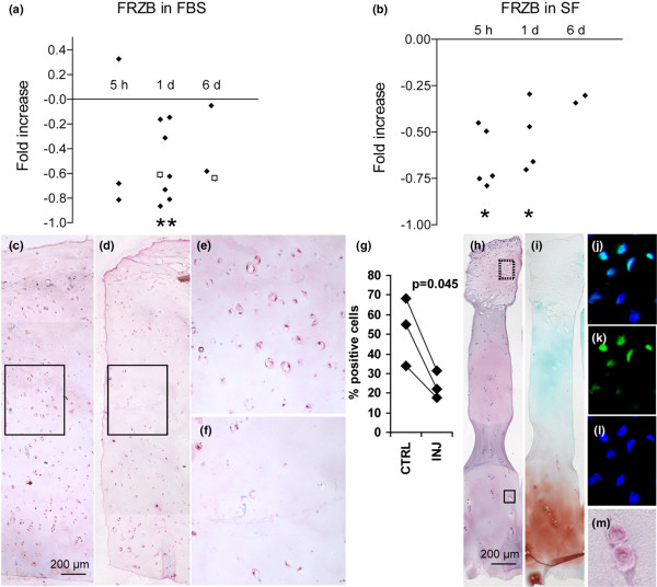
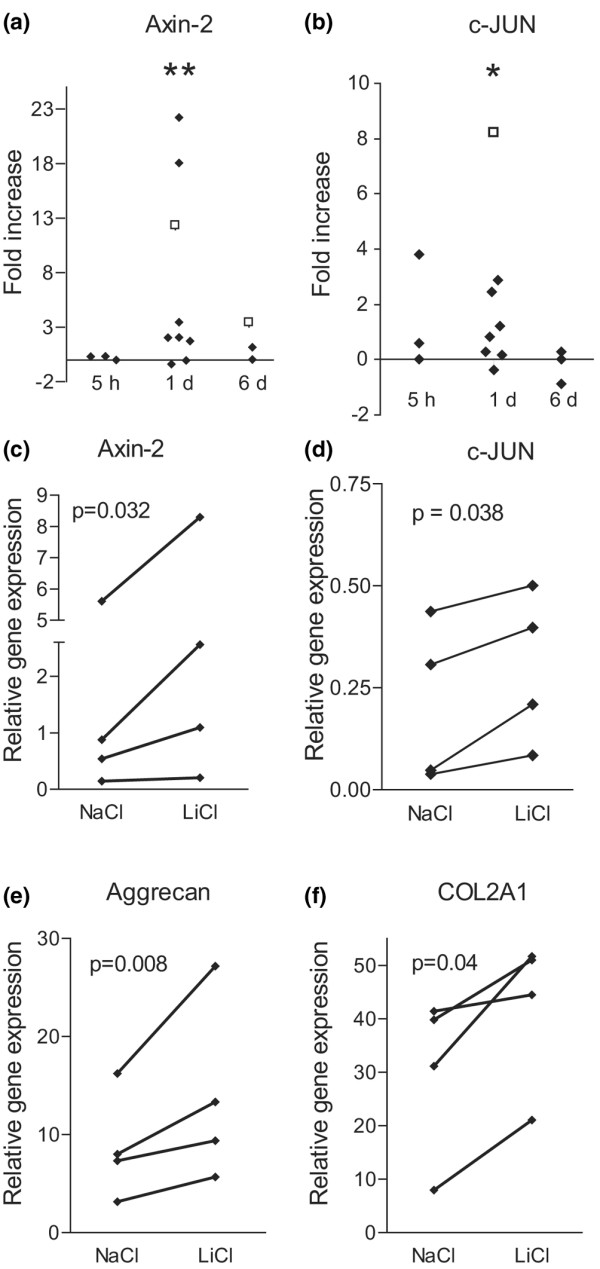
Acute full thickness joint surface defects can undergo repair, which involves tissue patterning and endochondral bone formation. Molecular signals regulating this process may contribute to the repair outcome, chronic evolution and, eventually, the onset of osteoarthritis. We tested the hypothesis that mechanical injury modulates morphogenetic pathways in adult human articular cartilage explants. Adjacent articular cartilage explants were obtained from preserved areas of the femoral condyles of patients undergoing arthroplasty for osteoarthritis, or from a normal joint of a patient undergoing lower limb amputation. Paired explants were individually maintained in explant culture. From each pair, one explant was mechanically injured and the other left uninjured as a control. Cultures were terminated at different time points for histochemistry, immunohistochemistry and gene expression analysis by reverse transcription real time PCR. Bone morphogenetic protein 2 (BMP-2) mRNA was upregulated in the injured explants. We detected phosphorylation of SMAD-1 and SMAD-5, consistent with activation of the bone morphogenetic protein (BMP) pathway. FRZB-1 mRNA was downregulated in the injured explants, suggesting de-repression of WNT signaling. Accordingly, expression of the canonical WNT target genes Axin-2 and c-JUN was upregulated in the injured explants. Activation of the canonical WNT signaling pathway by LiCl treatment induced upregulation of COL2A1 and Aggrecan mRNA, suggesting an anabolic effect. Phosphorylation of SMAD-1/-5 and downregulation of FRZB were confirmed in vivo in a mouse model of joint surface injury. Taken together, these data show modulation of the BMP and WNT pathways following mechanical injury in vitro and in vivo, which may play a role in the reparative response of the joint surface. These pathways may, therefore, represent potential targets in protocols of biological joint surface defect repair.


Figures

Figure 1
Figure 1
Ex vivo model of mechanical injury to adult human articular cartilage explants. (a) Adjacent explants from human adult articular cartilage were dissected and placed in culture in separate bacteriological Petri dishes. After 6 days, 1 explant was injured. At different time points the cultures were terminated for gene expression analysis, histochemistry and immunohistochemistry. (b) Safranin O staining of: a, freshly dissected normal articular cartilage; b, an adjacent explant after 7 days in culture; c, a further adjacent explant after 6 days in culture before injury plus 1 additional day after injury; and d, a typical freshly dissected explant from a preserved area from a patient who had undergone joint arthroplasty for osteoarthritis. (c,d) Time course of metalloproteinase (MMP)-3 and MMP-13 mRNA differential expression in injured versus uninjured explants. Values are normalized for the housekeeping gene β actin and expressed as fold change of gene expression in the injured explants from paired uninjured controls. Diamonds indicate samples from preserved areas from joints affected by osteoarthritis; open squares indicate sample pairs from healthy cartilage. *p < 0.05; **p < 0.01. D, day(s); h, hours.
Figure 2
Figure 2
Differential expression of bone morphogenetic protein (BMP)-2 and FRZB mRNA following mechanical injury. (a) BMP-2 mRNA was significantly upregulated and (b) FRZB mRNA significantly down-regulated in most injured samples compared to uninjured adjacent controls. Values were calculated using a standard curve and normalized for the housekeeping β actin gene. Diamonds indicate samples from preserved areas from joints affected by osteoarthritis; open squares indicate the sample pair from healthy cartilage.
Figure 3
Figure 3
Activation of the bone morphogenetic protein (BMP) signaling pathway. (a,b) Time course of the differential expression of BMP-2 mRNA in injured versus uninjured explants in (a) the presence or (b) the absence of fetal bovine serum (FBS) in the culture medium. Values are normalized for the housekeeping β actin gene and expressed as fold change of gene expression in the injured explants from paired uninjured controls. Diamonds indicate samples from preserved areas from joints affected by osteoarthritis; open squares indicate the sample pair from healthy cartilage. (c-g) Immunostaining for phosphorylated SMAD-1/-5 in: (c) freshly dissected normal cartilage; (g) the adjacent injured explant at day 1 after injury; (f) and the adjacent uninjured control at the same time-point. (d) Larger magnification of the area shown in the square in (c). In the freshly dissected sample, phosphorylated SMAD-1/-5-positive cells were detected predominantly in the intermediate layer indicated by the bracket in (c). (e) Image obtained by false coloring in red the image in (d) and superimposing it on the fluorescent image in the blue channel documenting the nuclear DAPI counterstain. The DAB precipitate in the phosphorylated SMAD-1/-5-positive cells quenched the DAPI fluorescence and, therefore, in this panel, phosphorylated SMAD-1/-5-positive cells appear red and the nuclei of negative cells appear blue. The top insets in (f,g) are large magnifications of the corresponding squared areas. (h) A graphic summary of the proportion of phospho-SMAD-1/-5-positive cells and the expression of BMP-2, FRZB, metalloproteinase (MMP)-3 and MMP-13 mRNAs in this experiment with normal adult human articular cartilage. Values are expressed as: percent of positive cells for phospho-SMAD-1/-5; relative gene expression normalized for the housekeeping β actin gene; percent of the day 6 time point for BMP-2, MMP-3 and MMP-13 mRNA; and percent of the freshly dissected cartilage for FRZB. *p < 0.05; **p < 0.01. D, day(s); H, hours; SF, serum free medium.
Figure 4
Figure 4
A figure showing modulation of the BMP and WNT pathway after mechanical injury in vivo in mice. Modulation of BMP and WNT pathway after mechanical injury in vivo in mice. 7 week old C57BL/6 male mice were challenged in a model of joint surface injury in vivo. In this model the knee joint surface is exposed by medial para-patellar arthrotomy and lateral patellar dislocation. A full thickness injury is made in the patellar groove using a custom made device in which the length of a 26G needle is limited by a glass bead (injured knee), or left uninjured (sham operated control). In either case the patellar dislocation is then reduced and the joint capsule and the skin sutured in separate layers and the mice allowed to walk freely. The animals were killed at different time-points for histological and histochemical analysis. A-B immunohistochemistry for FRZB in sham operated (A) and injured (B) articular cartilage 1 day after the operation. C-D immunohistochemistry for phosphorylated SMAD-1 in sham operated (A) and injured (B) articular cartilage 6 days after the operation. The asterisk indicates the site of injury (occupied by debris). The dashed line indicates the margin of the injury site.
Figure 5
Figure 5
Components of the canonical WNT pathway in adult human articular cartilage. (a,b) Time course of the differential expression of FRZB mRNA in injured versus uninjured explants in (a) the presence or (b) the absence of fetal bovine serum (FBS) in the culture medium. Values were calculated using a standard curve, normalized for the housekeeping β actin gene and expressed as fold change of gene expression in the injured explants from paired uninjured controls. Diamonds indicate samples from preserved areas from joints affected by osteoarthritis; open squares indicate sample pairs from healthy cartilage. (c-f) Immunohistochemical staining for FRZB protein (red) in (c) uninjured and (d) injured explants at the day 1 time point. Haematoxylin was used as a nuclear counterstain. (e,f) Larger magnifications of the boxed areas in (c) and (d), respectively. (g) Percentage of FRZB-positive cells in injured explants and in the paired uninjured controls from 3 independent donors as evaluated by immunohistochemistry. (h) Haematoxylin-eosin and (i) safranin O stainings of an explant with a relatively high degree of osteoarthritis (modified Mankin score 5). (j-m) Immunostaining for β catenin in parallel, non-consecutive sections of (h) and (i). (j-l) Indirect immunofluorescence stainings for β catenin from a parallel section in the area of (h) boxed with the dashed line (top). (k) β catenin (green). (l) DAPI counterstain of the same section (blue). (j) The superimposition of (k) and (l). In this tissue, which is commonly called pannus, there were cells with a nuclear localization of β catenin. (m) Immunohistochemistry showing the cytoplasmic localization of β catenin in chondrocytes of the basal layer (area in (h) boxed with a solid line). *p < 0.05; **p < 0.01. D, day(s); H, hours; SF, serum free Medium.
Figure 6
Figure 6
Activation of the WNT/β catenin canonical pathway following mechanical injury. (a) Axin-2 and (b) c-JUN mRNAs, two known transcriptional targets of the WNT/β catenin canonical pathway, were upregulated 1 day after injury compared to uninjured controls. (c-f) Paired cartilage explants were cultured in the presence of either 10 mM LiCl or 10 mM NaCl for 1 day and then terminated for gene expression analysis by quantitative real time PCR. Culture in the presence of LiCl induced the upregulation of axin-2 (c) and c-JUN (d) mRNAs, thereby confirming that these two genes are targets of the WNT/β catenin canonical pathway in this experimental system. LiCl treatment also upregulated aggrecan and COL2A1 mRNA (e,f). **p < 0.01. D, day(s); h, hours.

References

    1. Buckwalter JA, Saltzman C, Brown T, Schurman DJ. The impact of osteoarthritis: implications for research. Clin Orthop. 2004;(427 Suppl):S6–S15. - PubMed
    1. Messner K, Maletius W. The long-term prognosis for severe damage to weight-bearing cartilage in the knee: a 14-year clinical and radiographic follow-up in 28 young athletes. Acta Orthop Scand. 1996;67:165–168. - PubMed
    1. Shelbourne KD, Jari S, Gray T. Outcome of untreated traumatic articular cartilage defects of the knee: a natural history study. J Bone Joint Surg Am. 2003;85-A(Suppl 2):8–16. - PubMed
    1. Koshino T, Wada S, Ara Y, Saito T. Regeneration of degenerated articular cartilage after high tibial valgus osteotomy for medial compartmental osteoarthritis of the knee. Knee. 2003;10:229–236. doi: 10.1016/S0968-0160(03)00005-X. - DOI - PubMed
    1. Linden B. Osteochondritis dissecans of the femoral condyles: a long-term follow-up study. J Bone Joint Surg Am. 1977;59:769–776. - PubMed

Publication types

MeSH terms

Substances

LinkOut - more resources