. 2006 Aug 7:7:31.
doi: 10.1186/1471-2121-7-31.

TBLR1 regulates the expression of nuclear hormone receptor co-repressors

Affiliations

TBLR1 regulates the expression of nuclear hormone receptor co-repressors

Xin-Min Zhang et al. BMC Cell Biol. .

Abstract

Background: Transcription is regulated by a complex interaction of activators and repressors. The effectors of repression are large multimeric complexes which contain both the repressor proteins that bind to transcription factors and a number of co-repressors that actually mediate transcriptional silencing either by inhibiting the basal transcription machinery or by recruiting chromatin-modifying enzymes.

Results: TBLR1 [GenBank: NM024665] is a co-repressor of nuclear hormone transcription factors. A single highly conserved gene encodes a small family of protein molecules. Different isoforms are produced by differential exon utilization. Although the ORF of the predominant form contains only 1545 bp, the human gene occupies approximately 200 kb of genomic DNA on chromosome 3q and contains 16 exons. The genomic sequence overlaps with the putative DC42 [GenBank: NM030921] locus. The murine homologue is structurally similar and is also located on Chromosome 3. TBLR1 is closely related (79% homology at the mRNA level) to TBL1X and TBL1Y, which are located on Chromosomes X and Y. The expression of TBLR1 overlaps but is distinct from that of TBL1. An alternatively spliced form of TBLR1 has been demonstrated in human material and it too has an unique pattern of expression. TBLR1 and the homologous genes interact with proteins that regulate the nuclear hormone receptor family of transcription factors. In resting cells TBLR1 is primarily cytoplasmic but after perturbation the protein translocates to the nucleus. TBLR1 co-precipitates with SMRT, a co-repressor of nuclear hormone receptors, and co-precipitates in complexes immunoprecipitated by antiserum to HDAC3. Cells engineered to over express either TBLR1 or N- and C-terminal deletion variants, have elevated levels of endogenous N-CoR. Co-transfection of TBLR1 and SMRT results in increased expression of SMRT. This co-repressor undergoes ubiquitin-mediated degradation and we suggest that the stabilization of the co-repressors by TBLR1 occurs because of a novel mechanism that protects them from degradation. Transient over expression of TBLR1 produces growth arrest.

Conclusion: TBLR1 is a multifunctional co-repressor of transcription. The structure of this family of molecules is highly conserved and closely related co-repressors have been found in all eukaryotic organisms. Regulation of co-repressor expression and the consequent alterations in transcriptional silencing play an important role in the regulation of differentiation.


Figures

Figure 1
Figure 1
Comparison of the amino acid sequence of TBLR1 with other family members and with the homologous proteins in other species. The proteins shown in this alignment are: TBLR1-human.............................. NP078941, TBLR1-mouse............................... NP109657, TBL1X-human.............................. NP005638, TBL1Y-human............................... NP150600, TBL1-mouse.................................. XP135950, Ebi CG4063-PA [D. melanogaster] NP477329, A. gambia str. PEST....................... EA.A.12470, Arabidopsis; protein id: At5g........ NP201533, SIR4-interacting factor; SIF2p...... NP009661, Only proteins with homology to both the LisH domain (near the amino terminus) and the WD40 domains are included in the comparison. Regions of amino acid identity are shown in black; conservative substitutions are shown in gray. The large regions of non-homology are the result of insertions in the Arabidopsis and yeast sequences that have no counterparts in the other species.
Figure 2
Figure 2
Map of exons from TBLR1α and TBLR1β transcripts on the human chromosome 3q23 genomic sequence. The sequences in the exon marked with asterisks were not included in the original description of TBLR1 and are not included in NM024665.2. The first sequence [GenBank: AK022268] (*) maps on chromosome 3 between the first 2 exons of TBLR1. The second sequence [**] is DC42 [AF267864] and maps immediately 3' to TBLR1. The actual 3'end of the gene has not been experimentally defined. mRNA numbering begins with the first nucleotide identified at the 5' end of the sequence and the genomic sequence is numbered from the corresponding base in the DNA. The sequence begins with bp 12127476 of [GenBank: NT_005962.15].
Figure 3
Figure 3
Expression of TBLR1. a. Quantitative real time RT-PCR analysis of the expression of TBLR1, TBLR1β and TBL1 m RNA. The data shown are relative concentrations of mRNA normalized to the concentration of GAPDH mRNA found in each tissue. b. Correlation of TBLR1 & TBL1 Expression. The data shown were extracted from Figure 3a and compare the level of expression of TBLR1 and TBL1. c. Correlation of TBLR1 &TBLR1β Expression. The data shown were extracted from Figure 3a and compare the level of expression of total TBL1 and TBLR1β. d. Northern analysis. Blots of human total RNA (Multiple Tissue Northern Blot™) or blots prepared using RNA extracted from several human hematopoietic cell lines were analyzed with 32P labeled probes as shown in the figure. The same filter was used for all the tissue hybridizations. Between hybridizations the bound radioactive probes were stripped and the filter exposed to fresh film to assure that the stripping had been complete. The autoradiographs were scanned to produce the images shown.
Figure 4
Figure 4
a. Expression of TBLR1 in mouse tissues (Western Blotting). Anti peptide antibodies were made against deduced sequences from TBLR1. The antibody used was prepared against a sequence unique to TBL1 does not react with TBL1. After transfer, the membranes were stained with polyclonal rabbit anti-TBLR1 peptide antibodies and the transferred proteins visualized with HRP-labeled donkey anti-rabbit IgG. The bound HRP was detected using luminol as the substrate. b. Expression of TBLR1 by HK293T cells (Western Blotting). TBLR1 proteins were isolated by immuno-affinity chromatography from lysates of HK293T cells. Rabbit anti-TBLR1(117-125), covalently coupled to the gel, was used as the immunoadsorbant. The bound material was eluted with a glycine-HCl buffer (pH 2.8) and then run on the gels. After transfer to nitrocellulose, these were stained with either anti TLR1α or TRLR1β or with anti anti-TBLR1(117-125) which detects both isoforms. The proteins were visualized as above. c. Immunohistochemical Detection of the expression of TBLR1 in human fetal liver. All of the samples were from 17 week fetuses. The sample on the left was stained with the anti-TBLR1 antiserum that detects both isoforms but does not detect TBL1. The staining was detected with HRP coupled goat anti rabbit IgG using DAB as the chromagen. The original magnification was 40×. A digital image was captured and cropped in Photoshop. The images on the right show samples stained with the specific anti α and β sera. They were also detected with HRP coupled goat anti rabbit IgG but using AEC as a chromagen. The images were captured on film and then scanned to produce the digital images shown. The original magnification was 40×. The hematopoietic cells of the fetal liver stained more intensely with the anti TBLR1β antiserum. All of the samples were counterstained with haematoxylin.
Figure 5
Figure 5
Intracellular localization of TBLR1. 3T3 cells respond to apoptotic stimuli with increased expression of TBLR1 and with translocation of TBLR1 into the nucleus. grown in on collagen coated cover slips were used. Untreated cells in the log phase of growth are shown in panels a and d. Apoptosis was induced by either serum starvation (incubation with DMEM containing 0.2% FBS for 16 h) [panels b and e] or growth with100 ng/ml of the topoisomerase inhibitor, camptothecin (CPT) [panels d and f]. The top panels [a, b, and c] were stained for total TBLR1 with anti TBLR1 [117-125] while the bottom panels [d, e, and f] were stained with anti TBLR1β. The images were obtained using a Leitz Orthlux microscope at an original magnification of 600 × with a Nikon 60X phase fluorescence objective (NA 1.4) and captured with a Nikon digital camera.
Figure 6
Figure 6
Sequences required for the interaction of TBLR1 with SMRT. a. TBLR1 constructs were cloned in-frame into pCDNA3 with an N-terminal Flag tag. b. HuSMRT cDNA (1-900) was PCR-amplified and cloned in-frame into PGEX-6p-1. EcoR1 linearized plasmids were purified and used as a template for in vitro translation using 35S methionine. Expression of the GST fusion protein was induced with IPTG and the product adsorbed onto Glutathione sepharose 4B. For the pull-down experiment, 5 ng of 35S labeled and 20 ul of GST agarose were mixed. After incubation the bound material was eluted and electrophoresed through a 10% SDS PAGE gel. 10% of the labeled material was reserved and electrophoresed separately to demonstrate the efficiency of the original labeling.
Figure 7
Figure 7
Expression of TBLR1 alters the expression of SMRT and N-CoR. a. Transient expression of TBLR1 alters the expression of co-transfected SMRT(1-300). COS7 cells were co-transfected with pCDNA His-SMRT(1-300) and the His-tagged TBLR1 constructs described above. Control cells were transfected with pCDNA His-SMRT(1-300) and an empty vector.36 hrs after transfection the cells were lysed and equal amount of proteins were electrophoresed in SDS-PAGE gel. After transfer anti-His mAb was used to detect the transiently expressed proteins. The TBLR1 constructs used in this experiment were also His-tagged and are visible on the blot after staining. They are marked with * in the figure. The immuno-precipitates were quantified by phosphorimaging. After stripping, the blots were restained with anti ERK as a control for loading differences. b. Stable expression of TBLR1 alters the expression of endogenous N-CoR. Jurkat were infected with retroviruses expressing TBLR1 or the TBLR1 [δN] and TBLR1 [δC] construct. The retroviral vector also expressed GFP which was used to identify the infected cells. After two rounds of sorting the cells were analyzed by Western blotting for their expression of N-CoR. Th blot was then stripped and restained with an antibody to α tubulin as a loading control. c. Stable expression of TBLR1 does not alter the expression of endogenous N-CoR or SMRT message. mRNA was prepared from the cell lines used in Figure 3b and examined using quantitative real-time RT-PCR. Primer pairs that identified GAPDH and N-CoR were used. The results shown are representative of 3 experiments performed in triplicate. The samples are labeled as follows: black = control Jurkat; red = Jurkat (TBLR1); blue = Jurkat (TBLR1 [δC]); green = Jurkat(TBLR1 [δN])
Figure 8
Figure 8
Effect of proteasome inhibitors on SMRT (1-300) expression. COS7 cells, expressing His -SMRT(1-300) and GFP-His6 were treated with 15 um MG132 for 10 hours. The His-tagged proteins were detected by Western blotting.
Figure 9
Figure 9
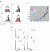
Growth inhibition after transfection with TBLR1. a. DiI staining of HK293T cells 72 hours after transfection with TBLR1 constructs. The cells were stained with DiI prior to transfection. The lipophilic dye distributes to both daughter cells after division and the loss of the dye reflects the number of times the cells have divided. The color filled curves are the computer generated model of the components of the mixed population. b. Growth of JURKAT cells over-expressing TBLR1. JURKAT cells infected with retroviral constructs expressing only GFP or GFP and a TBLR1 construct were seeded at a concentration of 25,000 per ml in 12 well Costar plates. Each day the entire contents of a well was removed and counted using a Coulter Model Zf cell counter. Each culture was performed in triplicate and the mean and S.D have been plotted. c. Cell cycle parameters (DNA content) of log phase JURKAT cells ectopically expressing TBLR1. Cells were harvested while in log phase of growth (72 hrs after plating at 50,000 cell per ml) and stained with PI as described. Fluorescence from PI bound to DNA was analyzed using a BD FACScan cytometer and the list mode data analyzed using ModFit (Verity Software) to calculate cell cycle parameters.

Similar articles

Cited by

References

    1. Darimont BD, Wagner RL, Apriletti JW, Stallcup MR, Kushner PJ, Baxter JD, Fletterick RJ, Yamamoto KR. Structure and specificity of nuclear receptor-coactivator interactions. Genes Dev. 1998;12:3343–3356. - PMC - PubMed
    1. Hu I, Lazar MA. Transcriptional Repression by Nuclear Hormone Receptors. Trends Endocrinol Metab. 2000;11:6–10. doi: 10.1016/S1043-2760(99)00215-5. - DOI - PubMed
    1. Chen JD, Evans RM. A transcriptional co-repressor that interacts with nuclear hormone receptors. Nature. 1995;377:454–457. doi: 10.1038/377454a0. - DOI - PubMed
    1. Horlein AJ, Na.a.r AM, Heinzel T, Torchia J, Gloss B, Kurokawa R, Ryan A, Kamei Y, Soderstrom M, Glass CK. Ligand-independent repression by the thyroid hormone receptor mediated by a nuclear receptor co-repressor. Nature. 1995;377:397–404. doi: 10.1038/377397a0. - DOI - PubMed
    1. Li J, Wang J, Nawaz Z, Liu JM, Qin J, Wong J. Both corepressor proteins SMRT and N-CoR exist in large protein complexes containing HDAC3. EMBO J. 2000;19:4342–4350. doi: 10.1093/emboj/19.16.4342. - DOI - PMC - PubMed

Publication types

MeSH terms

LinkOut - more resources