. 2006 Nov;26(21):7821-31.
doi: 10.1128/MCB.00548-06. Epub 2006 Aug 7.

The Drosophila inhibitor of apoptosis protein DIAP2 functions in innate immunity and is essential to resist gram-negative bacterial infection

Affiliations

The Drosophila inhibitor of apoptosis protein DIAP2 functions in innate immunity and is essential to resist gram-negative bacterial infection

François Leulier et al. Mol Cell Biol. 2006 Nov.

Abstract

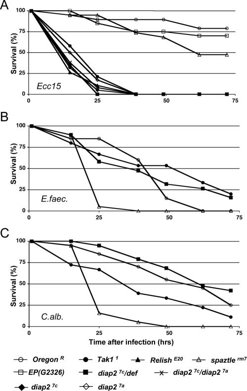
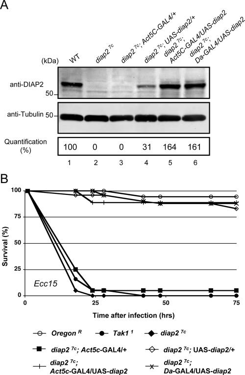
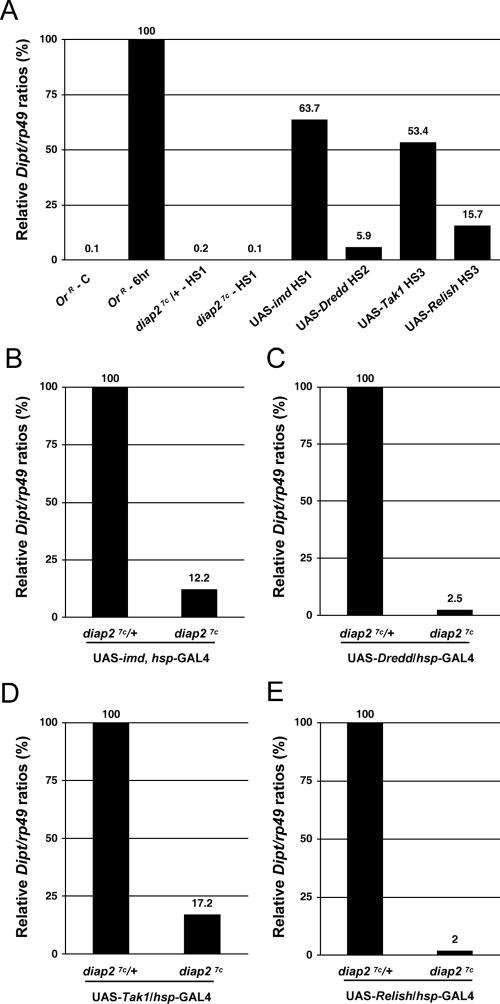
The founding member of the inhibitor of apoptosis protein (IAP) family was originally identified as a cell death inhibitor. However, recent evidence suggests that IAPs are multifunctional signaling devices that influence diverse biological processes. To investigate the in vivo function of Drosophila melanogaster IAP2, we have generated diap2 null alleles. diap2 mutant animals develop normally and are fully viable, suggesting that diap2 is dispensable for proper development. However, these animals were acutely sensitive to infection by gram-negative bacteria. In Drosophila, infection by gram-negative bacteria triggers the innate immune response by activating the immune deficiency (imd) signaling cascade, a NF-kappaB-dependent pathway that shares striking similarities with the pathway of mammalian tumor necrosis factor receptor 1 (TNFR1). diap2 mutant flies failed to activate NF-kappaB-mediated expression of antibacterial peptide genes and, consequently, rapidly succumbed to bacterial infection. Our genetic epistasis analysis places diap2 downstream of or in parallel to imd, Dredd, Tak1, and Relish. Therefore, DIAP2 functions in the host immune response to gram-negative bacteria. In contrast, we find that the Drosophila TNFR-associated factor (Traf) family member Traf2 is dispensable in resistance to gram-negative bacterial infection. Taken together, our genetic data identify DIAP2 as an essential component of the Imd signaling cascade, protecting the organism from infiltrating microbes.


Figures

FIG. 1.
FIG. 1.
Generation of DIAP2-deficient flies. (A) Imprecise excision of EP(G2326) created diap27c and diap27a alleles that carry deletions, which removed large portions of the diap2 locus. Schematic representation depicting the diap2 locus, the insertion site and orientation of EP(G2326), and the genomic DNA. The positions of the primers used to clone the respective genomic DNA fragments are indicated. (B) DIAP2 protein levels were undetectable in diap2 mutant flies. The presence of DIAP2 protein was examined by immunoblot analysis using anti-DIAP2 antibodies. Protein extracts from the following genotypes were used to monitor DIAP2 expression: diap27c/Cyo-actGFP (diap27c/+, third-instar larvae [L3]) (lane 1), diap27c (third-instar larvae) (lane 2), CantonS (wild type [WT], adult) (lane 3), EP(G2326) (adult) (lane 4), diap27c/Df(2R)exel7138 (diap27c/def, adult) (lane 5), diap27c (adult) (lane 6), diap27a (adult) (lane 7), or diap27a/diap27c (adult) (lane 8). Antitubulin immunoblot analysis was used to determine equal protein loading. (C) DIAP1 protein levels remain unchanged in diap2 mutant flies. The level of DIAP1 protein was examined by immunoblot analysis using anti-DIAP1 and antitubulin antibodies. Quantification of signals was performed using the LICOR system. Protein extracts from the following genotypes were analyzed: CantonS (WT, lane 1), diap27c (lane 2), diap27a (lane 3), or diap27a/diap27c (lane 4).
FIG. 2.
FIG. 2.
DIAP2 is required to resist gram-negative bacterial infection. The survival rates of adult males in response to different types of septic injuries are presented. Animals were pricked with a needle previously dipped into Erwinia carotovora subsp. carotovora 15 (Ecc15) (A), Enterococcus faecalis (E.faec.) (B), or Candida albicans (C.alb.) (C). The following genotypes were examined for susceptibility to microbes: wild-type (OregonR), Tak11, RelishE20, spatzlerm7, EP(G2326), diap27c/def, diap27c, diap27a, and diap27c/diap27a. Note that diap2 mutant flies behaved as Tak1 and Relish mutant flies, which are known to be highly susceptible to Erwinia carotovora subsp. carotovora 15 infection (A), but not to Enterococcus faecalis (B) or Candida albicans (C) infection. Both diap2 alleles, diap27c and diap27a, hemizygous (diap27c/def) or transheterozygous flies (diap27c/diap27a) showed similar susceptibility to E. carotovora subsp. carotovora 15 infection, while animals of the parental EP(G2326) line were fully resistant (A).
FIG. 3.
FIG. 3.
Ubiquitous expression of DIAP2 fully rescues the immune deficiency phenotype associated with diap27c. (A) Expression level of diap2 transgene in otherwise diap2 mutant flies. The presence of DIAP2 protein was examined by immunoblot analysis using anti-DIAP2 and antitubulin antibodies. Protein extracts from the following genotypes were used to monitor DIAP2 protein expression: CantonS (wild type [WT]) (lane 1), diap27c (lane 2), diap27c; Act5C-GAL4/+ (lane 3), diap27c; UAS-diap2/+ (lane 4), diap27c; UAS-diap2/Act5C-GAL4 (lane 5), and diap27c; UAS-diap2/Da-GAL4 (lane 6). Quantification of signals was performed using the LICOR system. (B) diap2 transgene expression rescued diap27c mutant flies from the lethal effects of Erwinia carotovora subsp. carotovora 15 (Ecc15)-mediated septic injury. The following genotypes were examined for susceptibility to microbes: wild type (OregonR), Tak11, diap27c, diap27c; Act5C-GAL4/+, diap27c; UAS-diap2/+, diap27c; Act5c-GAL4/UAS-diap2, and diap27c; Da-GAL4/UAS-diap2.
FIG. 4.
FIG. 4.
diap2 mutant individuals fail to induce antibacterial peptide genes following Erwinia carotovora subsp. carotovora 15 infection. (A and C) Quantitative RT-PCR analysis of Diptericin (Dipt) induction after E. carotovora subsp. carotovora 15 septic injury (Ecc15 SI) in diap27c, diap27c/def, Tak11, and RelishE20 mutants, diap27c; UAS-diap2/+ and diap27c; UAS-diap2/Da-GAL4 flies, and wild-type (OregonR) adult males. Results are shown for control (unchallenged) (C) flies and flies 6 and 24 hours after infection. (B) Drosomycin (Drs) induction after Micrococcus luteus septic injury (M.lut. SI) of wild-type (OregonR), diap27c/def, Tak11, and spatzlerm7 adult males. Results are shown for control (unchallenged) (C) flies and flies 24 and 48 hours after infection. (D) Attacin-A (AttA), Cecropin-A1 (CecA1), Defensin (Def), Drosocin (Dro), and Metchnikowin (Mtk) induction 24 h after E. carotovora subsp. carotovora 15 septic injury of wild-type (OregonR), diap27c/def, Tak11, and RelishE20 adult males. Similar to Tak1 and Relish mutants, diap2 mutant flies were significantly impaired in their ability to induce antibacterial peptide genes in response to gram-negative bacterial septic injury. However, these mutants showed normal Drosomycin induction following exposure to gram-positive bacteria. Diptericin induction after E. carotovora subsp. carotovora 15 infection was restored in diap27c mutant flies expressing the UAS-diap2 transgene. rp49 was used as the experimental expression standard. Shown are the relative expression ratios of Dipt/rp49 (A and C), Drs/rp49 (B), AttA/rp49, CecA1/rp49, Def/rp49, Dro/rp49, and Mtk/rp49 (D).
FIG. 5.
FIG. 5.
DIAP2 is required to mount a systemic antibacterial immune response to oral infection by Erwinia carotovora subsp. carotovora (Ecc15). Quantitative RT-PCR analysis of Diptericin (Dipt) (A), Drosocin (Dro) (B), and Attacin-A (AttA) (C) induction after E. carotovora subsp. carotovora 15 natural infection (Ecc15 NI) in wild-type (OregonR), diap27c/def, Tak11, and RelishE20 mutant third-instar larvae. Larvae were orally infected by exposing animals to food contaminated with E. carotovora subsp. carotovora 15. Similar to Tak1 and Relish mutants, diap2 mutant individuals failed to significantly induce Attacin-A, Diptericin, and Drosocin expression following natural infection. rp49 was used as an experimental expression standard. The relative Dipt/rp49 (A), Dro/rp49 (B), and AttA/rp49 (C) expression values for control (noninfected) (C) flies and flies 24 h after feeding are shown.
FIG. 6.
FIG. 6.
DIAP2 functions genetically downstream of or in parallel to imd, Dredd, Tak1, and Relish. Quantitative RT-PCR analysis of Diptericin expression following overexpression of imd, Dredd, Tak1, and Relish was performed. (A) Diptericin (Dipt) expression levels of the following animals are shown: control OregonR animals that were not challenged (OrR - C) and 6 h after Erwinia carotovora subsp. carotovora 15 septic injury (OrR - 6hr); diap27c/+ and diap27c mutant flies 3 h after 1 h of heat shock at 37°C (diap27c/+-HS1 and diap27c-HS1) or heat shock-mediated overexpression of imd (UAS-imd HS1), Dredd (UAS-Dredd HS2), Tak1 (UAS-Tak1 HS3), and Relish (UAS-Relish HS3). (B) In the absence of infection, heat shock-mediated imd overexpression caused high levels of Diptericin (Dipt) expression (63.7% of the level of Diptericin observed 6 h after E. carotovora subsp. carotovora 15 septic injury in panel A) that was significantly thwarted in diap27c mutant flies (87.8% reduction). (C) Heat shock-mediated Dredd overexpression triggered weak but reproducible Diptericin expression (5.9% of the levels of Diptericin observed after 6 h of E. carotovora subsp. carotovora 15 septic injury in panel A), which was blocked in diap27c mutant flies. (D) Heat shock-mediated Tak1 overexpression triggered strong Diptericin expression (53.4% of Diptericin levels observed after 6 h of E. carotovora subsp. carotovora 15 septic injury in panel A) which was blocked in diap27c mutants. (E) Heat shock-mediated Relish overexpression triggered weak but reproducible Diptericin expression (15.7% of the levels of Diptericin observed after 6 h of E. carotovora subsp. carotovora 15 septic injury in panel A) which was not observed in diap27c mutant flies. Flies were heat shocked for 1 hour at 37°C and left to recover at 25°C for 3 h (A and B) (HS1), 1 h (A and C) (HS2), or 24 h (A, D, and E) (HS3) prior to analysis. The analyzed genotypes were as follows: (i) diap27c/Cyo, act-GFP (diap27c/+), diap27c; (ii) diap27c/Cyo; UAS-imd, hsp-GAL4/TM6Tb (UAS-imd), diap27c; UAS-imd, hsp-GAL4/TM6Tb; (iii) diap27c/Cyo; UAS-Dredd/hsp-GAL4 (UAS-Dredd), diap27c; UAS-Dredd/hsp-GAL4; (iv) diap27c, UAS-Tak1/+; hsp-GAL4/+ (UAS-Tak1), diap27c, UAS-Tak1/diap27c; hsp-GAL4/+; (v) diap27c, UAS-Relish/+; hsp-GAL4/+ (UAS-Relish); (vi) diap27c, UAS-Relish/diap27c; hsp-GAL4/+.
FIG. 7.
FIG. 7.
Traf2 is dispensable in resisting gram-negative bacterial infection. Shown are the survival rates of Traf2 null mutant adult males exposed to septic injury with E. carotovora subsp. carotovora 15 (Ecc15). The following genotypes were examined: wild type (OregonR), Tak11, diap27c, and Traf2ex1. Note that Traf2-deficient flies behaved like wild-type flies, while diap2 and Tak1 mutant flies were highly susceptible to E. carotovora subsp. carotovora 15-mediated septic injury.
FIG. 8.
FIG. 8.
Model of Imd signaling based on genetic epistasis data in vivo. Our genetic epistasis analysis places diap2 downstream of or in parallel to imd, Dredd, Tak1, and Relish. Intriguingly, loss of diap2 copies the phenotype of Tak1 mutant animals, since Diptericin induction, following enforced expression of imd and Dredd, is also blocked in Tak1 mutant animals, while this is not the case in kenny and ird5 mutant flies. The Imd signal transduction pathway bifurcates at the level of Tak1, which is required for the activation of the NF-κB signaling branch as well as the JNK signaling branch, both of which are necessary for expression of antibacterial peptide genes in the fat body. We currently favor the model whereby diap2 functions genetically at the level of Tak1. This view is supported by recent reports of Drosophila tissue culture cells which suggest that DIAP2 is required for Tak1-mediated JNK activation. Arrows indicate genetic interactions that rely on overexpression of individual components of the Imd pathway in vivo. Note that the ability to induce Diptericin expression varied substantially among heat shock-induced overexpression of imd, Dredd, Tak1, and Relish (see Fig. 6 for more details). AMPs, antimicrobial peptides.

References

    1. Basset, A., R. Khush, A. Braun, L. Gardan, F. Boccard, J. Hoffmann, and B. Lemaitre. 2000. The phytopathogenic bacteria Erwinia carotovora infects Drosophila and activates an immune response. Proc. Natl. Acad. Sci. USA 97:3376-3381. - PMC - PubMed
    1. Cha, G. H., K. S. Cho, J. H. Lee, M. Kim, E. Kim, J. Park, S. B. Lee, and J. Chung. 2003. Discrete functions of TRAF1 and TRAF2 in Drosophila melanogaster mediated by c-Jun N-terminal kinase and NF-κB-dependent signaling pathways. Mol. Cell. Biol. 23:7982-7991. - PMC - PubMed
    1. Chen, Z. J., V. Bhoj, and R. B. Seth. 2006. Ubiquitin, TAK1 and IKK: is there a connection? Cell Death Differ. 13:687-692. - PubMed
    1. Choe, K. M., H. Lee, and K. V. Anderson. 2005. Drosophila peptidoglycan recognition protein LC (PGRP-LC) acts as a signal-transducing innate immune receptor. Proc. Natl. Acad. Sci. USA 102:1122-1126. - PMC - PubMed
    1. Conte, D., M. Holcik, C. A. Lefebvre, E. Lacasse, D. J. Picketts, K. E. Wright, and R. G. Korneluk. 2006. Inhibitor of apoptosis protein cIAP2 is essential for lipopolysaccharide-induced macrophage survival. Mol. Cell. Biol. 26:699-708. - PMC - PubMed

Publication types

MeSH terms