. 2006 Aug 15;103(33):12411-6.
doi: 10.1073/pnas.0602443103. Epub 2006 Aug 7.

Myosin-X is a molecular motor that functions in filopodia formation

Affiliations

Myosin-X is a molecular motor that functions in filopodia formation

Aparna B Bohil et al. Proc Natl Acad Sci U S A. .

Abstract

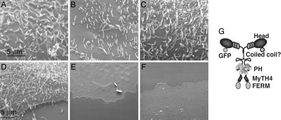
Despite recent progress in understanding lamellipodia extension, the molecular mechanisms regulating filopodia formation remain largely unknown. Myo10 is a MyTH4-FERM myosin that localizes to the tips of filopodia and is hypothesized to function in filopodia formation. To determine whether endogenous Myo10 is required for filopodia formation, we have used scanning EM to assay the numerous filopodia normally present on the dorsal surfaces of HeLa cells. We show here that siRNA-mediated knockdown of Myo10 in HeLa cells leads to a dramatic loss of dorsal filopodia. Overexpressing the coiled coil region from Myo10 as a dominant- negative also leads to a loss of dorsal filopodia, thus providing independent evidence that Myo10 functions in filopodia formation. We also show that expressing Myo10 in COS-7 cells, a cell line that normally lacks dorsal filopodia, leads to a massive induction of dorsal filopodia. Because the dorsal filopodia induced by Myo10 are not attached to the substrate, Myo10 can promote filopodia by a mechanism that is independent of substrate attachment. Consistent with this observation, a Myo10 construct that lacks the FERM domain, the region that binds to integrin, retains the ability to induce dorsal filopodia. Deletion of the MyTH4-FERM region, however, completely abolishes Myo10's filopodia-promoting activity, as does deletion of the motor domain. Additional experiments on the mechanism of Myo10 action indicate that it acts downstream of Cdc42 and can promote filopodia in the absence of VASP proteins. Together, these data demonstrate that Myo10 is a molecular motor that functions in filopodia formation.


Conflict of interest statement

Conflict of interest statement: No conflicts declared.

Figures

Fig. 1.
Fig. 1.
Expressing Myo10 leads to a massive increase in dorsal filopodia. (A and B) The dorsal surfaces of control COS-7 cells transfected with GFP alone are smooth and lack dorsal filopodia. (C and D) Cells transfected with GFP-Myo10, however, exhibit a massive increase in dorsal filopodia. A few substrate-attached filopodia also are visible. (EG) Fluorescence microscopy demonstrates that the dorsal structures induced by GFP-Myo10 contain known markers of filopodia, including F-actin (E) and fascin (F), which label filopodial shafts, and VASP (G), which is present in filopodial tips. Note that GFP-Myo10 is visible at the tips of the numerous dorsal filopodia induced in these cells and, in some cases, forms streaks extending into the filopodial shaft. Although the SEM images in this figure were obtained from an experiment where FACS was used to isolate transfected cells before SEM, similar results were obtained when fluorescence-correlative SEM was used to image transfected cells (Figs. 6 AC and 7A, which are published as supporting information on the PNAS web site).
Fig. 2.
Fig. 2.
Expressing VASP, fascin, Cdc42, or a Myo10 construct lacking the FERM domain also lead to massive increases in dorsal filopodia. (AC) Transfecting known inducers of filopodia such as GFP-VASP (A), GFP-fascin (B), and constitutively active GFP-Cdc42(61L) (C) in COS-7 cells all lead to massive increases in dorsal filopodia. Note that in addition to filopodia, the VASP and Cdc42 constructs sometimes also induced small ruffle-like structures. (DF) Domain-mapping experiments show that GFP-Myo10 lacking the FERM domain retains the ability to induce dorsal filopodia when expressed in COS-7 cells, whereas GFP-Myo10 lacking the MyTH4 (E) and FERM (D) domains fails to induce filopodia. The GFP-Myo10-heavy meromyosin construct, which lacks the PH, MyTH4, and FERM (F) domains, also fails to induce filopodia. (G) A hypothetical model of Myo10 as a dimer illustrating its major domains. The SEM images illustrated here and in all subsequent figures were obtained by using fluorescence-correlative SEM.
Fig. 3.
Fig. 3.
Inhibiting Myo10 suppresses dorsal filopodia. (A) Immunoblot of HeLa cells treated with control or Myo10 siRNA showing specific knockdown of Myo10. Samples were run with a 4–20% SDS/PAGE, transferred to nitrocellulose, and stained with Ponceau to reveal total protein and then blotted with anti-Myo10 to confirm knockdown. (B) SEM of a HeLa cell from the same experiment treated with control siRNA showing the numerous dorsal filopodia normally present on these cells. (C) SEM of a HeLa cell from the same experiment treated with Myo10 siRNA showing the loss of dorsal filopodia induced by Myo10 siRNA. (D) SEM of a HeLa cell illustrating the loss of dorsal filopodia observed in HeLa cells expressing the dominant-negative GFP-Myo10 coiled-coil construct.
Fig. 4.
Fig. 4.
Myo10 acts downstream of Cdc42. (A) Like untreated HeLa cells, HeLa cells treated with a control siRNA and then transfected with constitutively active GFP-Cdc42 exhibit numerous dorsal filopodia. (B) Parallel samples treated with the Myo10 siRNA, however, have very few dorsal filopodia, even in the presence of constitutively active GFP-Cdc42, indicating that Myo10 functions downstream of Cdc42. (C and D) The loss of dorsal filopodia induced by the siRNA to human Myo10 (C) can be rescued by transfection with bovine GFP-Myo10 (D). Note that the constitutively active GFP-Cdc42(61L) construct used here led to a massive induction of filopodia in other situations (Fig. 2C).
Fig. 5.
Fig. 5.
Myo10 can induce dorsal filopodia in the absence of VASP family proteins. (A) Ena/VASP null cells (MVD7) transfected with GFP alone exhibit very few dorsal filopodia. (B) Ena/VASP null cells transfected with GFP-Myo10, however, exhibit numerous dorsal filopodia, thus demonstrating that Myo10 does not require VASP proteins for its filopodia promoting activity.

References

    1. O’Connor T. P., Duerr J. S., Bentley D. J. Neurosci. 1990;10:3935–3946. - PMC - PubMed
    1. Wood W., Jacinto A., Grose R., Woolner S., Gale J., Wilson C., Martin P. Nat. Cell Biol. 2002;4:907–912. - PubMed
    1. Gerhardt H., Golding M., Fruttiger M., Ruhrberg C., Lundkvist A., Abramsson A., Jeltsch M., Mitchell C., Alitalo K., Shima D., Betsholtz C. J. Cell Biol. 2003;161:1163–1177. - PMC - PubMed
    1. Ramirez-Weber F. A., Kornberg T. B. Cell. 2000;103:189–192. - PubMed
    1. Pollard T. D., Borisy G. G. Cell. 2003;112:453–465. - PubMed

Publication types

MeSH terms