. 2006 Sep;235(9):2507-20.
doi: 10.1002/dvdy.20891.

Endogenous bone morphogenetic protein antagonists regulate mammalian neural crest generation and survival

Affiliations

Endogenous bone morphogenetic protein antagonists regulate mammalian neural crest generation and survival

Ryan M Anderson et al. Dev Dyn. 2006 Sep.

Abstract

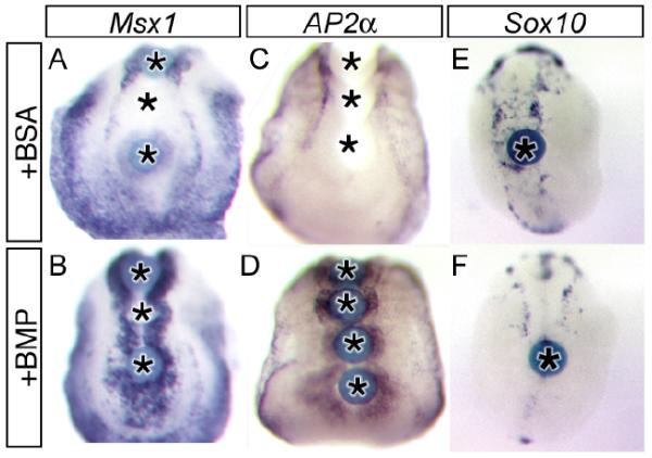
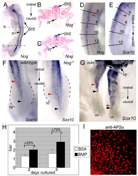
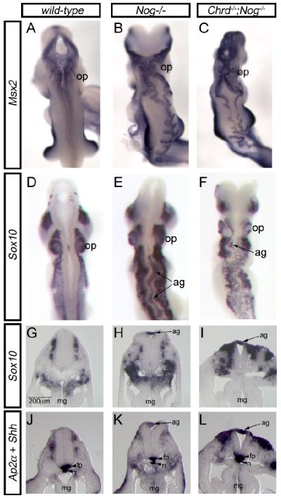
We demonstrate here that Chordin and Noggin function as bone morphogenetic protein (BMP) antagonists in vivo to promote mammalian neural crest development. Using Chrd and Nog single and compound mutants, we find that Noggin has a major role in promoting neural crest formation, in which Chordin is partially redundant. BMP signaling is increased in dorsal tissues lacking Noggin and is further increased when Chordin is also absent. The early neural crest domain is expanded with decreased BMP antagonism in vivo. Noggin and Chordin also regulate subsequent neural crest cell emigration from the neural tube. However, reduced levels of these BMP antagonists ultimately result in perturbation of neural crest cell derived peripheral nervous system and craniofacial skeletal elements. Such defects reflect, at least in part, a function to limit apoptosis in neural crest cells. Noggin and Chordin, therefore, function together to regulate both the generation and survival of neural crest cells in mammalian development.


Figures

Figure 1
Figure 1
Increased BMP signaling and increased neural crest cell generation in Nog−/− and Chrd−/−;Nog−/− mutants. (A-C) Msx2 expression used as a measure of BMP signaling in the dorsal neural tube. Compared to wild-type 8-9 somite embryos (A), increased Msx2 expression is observed in the dorsal neural tube of Nog−/− (B) and Chrd−/−;Nog−/− (C) mutants at all axial levels. In Chrd−/−;Nog−/− mutants, Msx2 expression is increased in the lateral plate mesoderm (lpm) as well. (D-F) Ap2α expression at the neural epidermal boundary and in neural crest cells. Compared to wild-type 6-7 somite embryos (D), expression is increased in Nog−/− (E) and Chrd−/−;Nog−/− (F) mutants. (G-L) Sox10 expression in neural crest cells. At the 8-9 somite stage, Sox10 expression is unchanged in Nog−/− mutants (H), and slightly expanded in Chrd−/−;Nog−/− mutants (I). At the 11-12 somite stage, expression is dramatically increased in both Nog−/− (K) and Chrd−/−;Nog−/− (L) mutant embryos. This region of expanded Sox10 expression in the trunk is indicated by brackets. Abbreviations: mb, midbrain; hb, hindbrain; tr, trunk; cn, caudal neuropore.
Figure 2
Figure 2
BMP is not sufficient for neural crest cell induction in vitro. In wild-type explants of 5-7 somite embryos cultured for 7 hours with protein-soaked beads, BMP2, but not BSA, elicited increased amounts of Msx1 (B) and Ap2α Bone morphogenetic protein (BMP) is not sufficient for neural crest cell induction in vitro. In wild-type explants of five- to seven-somite embryos cultured for 7 hr with protein-soaked beads, BMP2, but not bovine serum albumin (BSA), elicited increased amounts of Msx1 (B) and Ap2α adjacent to protein-soaked beads. adjacent to protein-soaked beads. Increased Sox10 expression (E, F) was not observed with either treatment. Indeed, Sox10 expression sometimes was decreased locally around the BMP beads (F), possibly due to increased cell death of NCC derivatives (see text). Bead locations during culture are indicated by asterisks.
Figure 3
Figure 3
Precocious delamination of NCCs in Nog−/− and Chrd−/−;Nog−/− embryos. (A-D) Increasing NCC delamination (rostral to caudal) is reciprocal to Nog expression gradient. (A) Graded Nog expression in dorsal neural tube (dnt) of 10-somite embryo as detected by in situ hybridization. Expression in notochord (n) is not graded, and serves as an internal control. Rostral (B) and caudal (C) sections of embryo shown in panel A. Dorsal neural tube expression is stronger at more caudal levels. (D) In ~12s embryos, Nog expression is diminished rostrally, beginning at the level of the 6th–8th somite pair (red arrowheads). (E) Sox10 expression is observed in NCCs emigrating from the dorsal midline beginning at the axial levels where Nog expression is diminished in the neural tube. (F) Dorsal aspect of trunk region of 22s wild-type and Nog−/− mutant embryos labeled with Sox10 to mark NCCs. Both non-migrating (black arrows) and migrating NCCs (red arrows) are seen further caudally in Nog−/− mutants than in wild-type littermates (compare to axial level of hindlimb, hl, outlined by dashed lines). (G) Sox10 expression in wild-type and Chrd−/−;Nog−/− littermates. Axial levels of NCC specification (black arrows) and migration (red arrows) are displaced further caudally in mutants (compare distance to closing lip of caudal neuropore). Exact stage matching by somite counting was not possible due to aberrant somite development in Chrd−/−;Nog−/− mutants. (H) Treatment of cultures with recombinant BMP2 (n = 32) increased the migration index of neural crest cells by 54.4% after 2 days of culture, and by 79.4% after 3 days of culture, relative to explants cultured in BSA-treated medium (n = 18) (p <0.001 for both comparisons). BMP concentrations of 100ng/ml and 200ng/ml gave results with no statistically significant difference, and were therefore pooled. (I) Antibody staining of NCC outgrowth cultures for AP2α. All migratory cells from trunk explants express AP2α, All migratory cells from trunk explants express AP2α, indicating their identity as NCCs. Core of tissue at the lower left is a neural tube explant.
Figure 4
Figure 4
BMP signaling and neural crest phenotypes at E9.5. Wild-type (A,D,G,J), Nog−/− mutant (B,E,H,K), and Chrd−/−;Nog−/− (C,F,I,L) mutant embryos. (A-C) Msx2 expression as and indicator of BMP signaling in the dorsal neural tube. (B) Nog−/− and (C) Chrd−/−;Nog−/− mutant embryos display increased Msx2 expression the dorsal midline. (D-F) Sox10 expression in NCCs. (E) Nog−/− and (F) Chrd−/−;Nog−/− mutants display an increase in dorsal NCCs; in both classes, ectopic aggregates (ag) of NCCs accumulate at the dorsal midline. (G-L) Transverse section in situ hybridization for Sox10 (G-I) and Ap2α +Shh (J-L) at the forelimb level. Increased amounts of NCCs are seen in (H,K) Nog−/− and (I,L) Chrd−/−;Nog−/− mutants. Distribution of NCCs in Chrd−/−;Nog−/− mutants is biased more proximally to the dorsal neural tube than in Nog−/− mutants. For example, note in Nog−/− mutants more Ap2α and Sox10 positive cells around the midgut (mg), whereas in Chrd−/−;Nog−/− more labeled cells are located dorsally. Ectopic aggregates are observed superficial to the dysmorphic dorsal neural tube of both mutant classes.
Figure 5
Figure 5
Deficiency of skeletal neural crest derivatives in Chrd−/−;Nog+/− mutants. Lateral (A-C) and ventral (D-G) views of wild-type (A,D) and Chrd−/−;Nog+/− mutant (B,C,E-G) skulls. Affected mutants have a variable skull phenotype (arranged left to right with increasing severity) and show a corresponding lack of neural crest-derived craniofacial skeletal elements, such as upper incisors (ui; position marked by asterisk in mutants), medial premaxillae (px) and palate, and presphenoid (ps). Lateral structures such as the tympanic rings (tr) zygomatic arches (za), maxillae (mx), and alisphenoids (al) are displaced medially (tympanic rings removed from wild-type for clarity; compare E and F). Other abbreviations: nasal, n; frontal, f; parietal, p; interparietal, ip; supraoccipital, so; otic capsule, oc; mandible, md; palatine, pl; basoccipital, bo; basosphenoid, bs.
Figure 6
Figure 6
Aberrant peripheral nervous system development with decreased BMP antagonism. (A-C) Lateral aspect of fluorescent neurofilament staining in E10.5 mutant embryos. Wild-type (A). In Nog−/− mutants (B) the vagus (X) is expanded and is indistinct from glossopharyngeal (IX). Severe Chrd−/−;Nog−/− mutants (C) lack most neurofilament immunoreactivity, but a rudimentary portion of the trigeminal ganglion (V) remains. (D-F) Higher magnification of dorsal root ganglia from mutant embryos. Wild-type (D). In Nog−/− mutants (E) DRGs are present, but are disarrayed and indistinct. In mild Chrd−/−;Nog−/− mutants (F), most DRGs are absent. Note neurofilament-positive aggregation (ag) dorsal to neural tube.
Figure 7
Figure 7
Ectopic BMP disrupts dorsal root ganglia formation in explants. E9.5 trunk explants cultured for 24 hours with beads incubated in BSA (top row) or BMP (bottom row). Asterisks indicate bead positions during culture. The left side of each explant serves as an internal control. (A) Msx1 expression at the dorsal midline. Msx1 expression is not elicited by BSA-coated beads, but is induced to high levels by BMP beads. (B) Sox10 expression in NCCs contributing to the peripheral nervous system. The distribution of Sox10-expressing cells is not affected by BSA beads, but is strongly disrupted by BMP2 beads. Arrows point to examples of dorsal root ganglia (drg). (C) NCC-derived sensory neurons stained for neurofilament (2H3) are not disrupted by BSA beads, but are significantly disrupted by BMP4-coated beads. (D) Genetic NCC-lineage tracing in explants prepared from Wnt1-Cre;R26R embryos; NCCs are stained blue. BSA beads has no effect, but BMP beads disrupts NCC contribution to peripheral tissue.
Figure 8
Figure 8
Chrd and Nog protect migratory neural crest cells from BMP-mediated apoptosis. Transverse sections of 11-12s wild-type (A), Nog−/− mutant (B), and Chrd−/−;Nog−/− mutant (C) embryos, taken at the level of pharyngeal arch 2. (A’-C’) Higher magnification of corresponding samples shown in A-C, as indicated by hatched boxes. Apoptotic TUNEL-positive cells are indicated in green, proliferating phospho-histone H3-positive cells in blue, and AP2α-positive cells in red, as in figure key (inset, A’). The number of NCCs and extent of NCC apoptosis (yellow, examples indicated by arrows) is increased in Nog−/− mutants, and much more so in Chrd−/−;Nog−/− mutants. Additionally, Chrd−/−;Nog−/− mutants show ectopic domains of AP2α-positve cells in the neural tube and streaming into the periphery (C, arrowheads). Differences in cell proliferation rates were not apparent at this stage. Positive signal in lumen of pharynx is specific staining of non-cellular material. Abbreviations: ot, otic placode, ph, pharynx.

References

    1. Abzhanov A, Protas M, Grant BR, Grant PR, Tabin CJ. Bmp4 and morphological variation of beaks in Darwin’s finches. Science. 2004;305:1464–1465. - PubMed
    1. Abu-Issa R, Smyth G, Smoak I, Yamamura K, Meyers EN. Fgf8 is required for pharyngeal arch and cardiovascular development in the mouse. Development. 2002;129:4613–25. - PubMed
    1. Ahlgren SC, Bronner-Fraser M. Inhibition of sonic hedgehog signaling in vivo results in craniofacial neural crest cell death. Curr Biol. 1999;9:1304–14. - PubMed
    1. Anderson RM, Lawrence AR, Stottmann RW, Bachiller D, Klingensmith J. Chordin and noggin promote organizing centers of forebrain development in the mouse. Development. 2002;129:4975–87. - PubMed
    1. Arkell R, Beddington RSP. BMP-7 influences pattern and growth of the developing hindbrain of mouse embryos. Devlopment. 1997;124:1–12. - PubMed

Publication types

MeSH terms

LinkOut - more resources