. 2006 Jul;4(8):e261.
doi: 10.1371/journal.pbio.0040261.

Tomosyn inhibits synaptic vesicle priming in Caenorhabditis elegans

Affiliations

Tomosyn inhibits synaptic vesicle priming in Caenorhabditis elegans

Elena O Gracheva et al. PLoS Biol. 2006 Jul.

Abstract

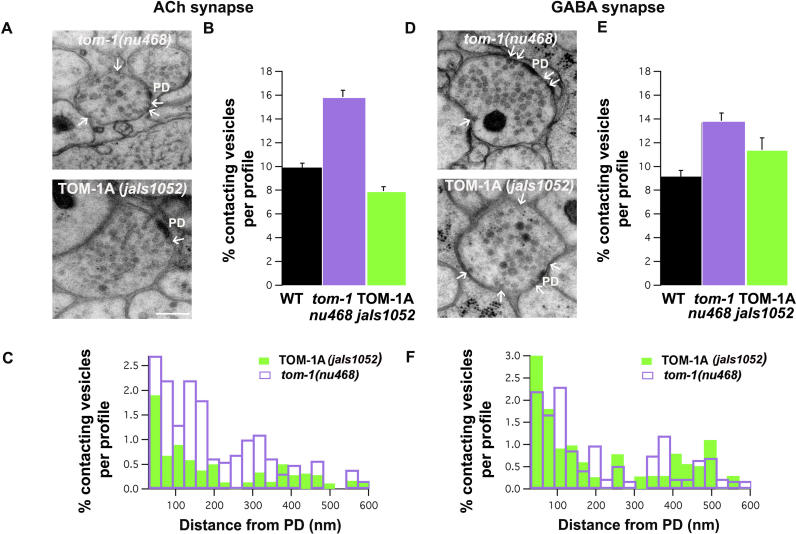
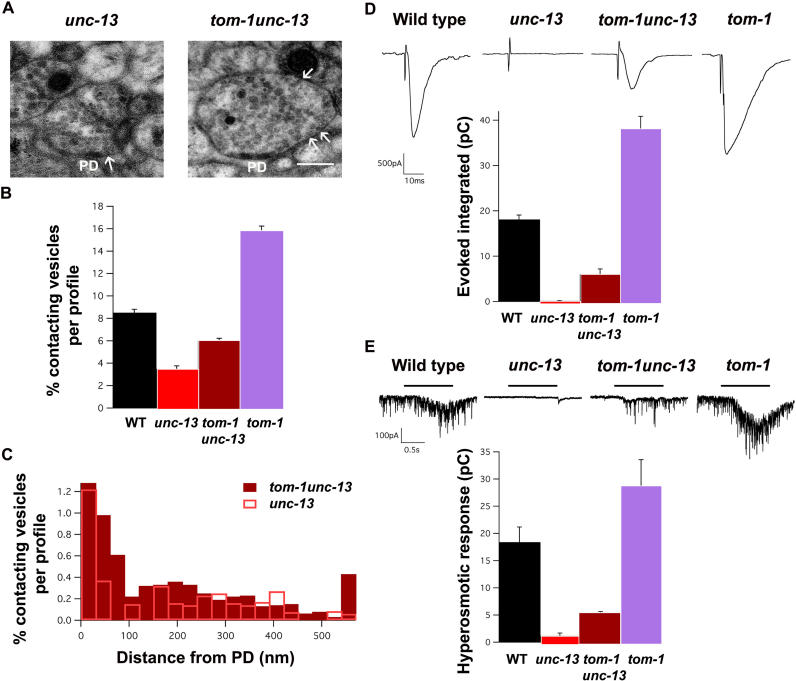
Caenorhabditis elegans TOM-1 is orthologous to vertebrate tomosyn, a cytosolic syntaxin-binding protein implicated in the modulation of both constitutive and regulated exocytosis. To investigate how TOM-1 regulates exocytosis of synaptic vesicles in vivo, we analyzed C. elegans tom-1 mutants. Our electrophysiological analysis indicates that evoked postsynaptic responses at tom-1 mutant synapses are prolonged leading to a two-fold increase in total charge transfer. The enhanced response in tom-1 mutants is not associated with any detectable changes in postsynaptic response kinetics, neuronal outgrowth, or synaptogenesis. However, at the ultrastructural level, we observe a concomitant increase in the number of plasma membrane-contacting vesicles in tom-1 mutant synapses, a phenotype reversed by neuronal expression of TOM-1. Priming defective unc-13 mutants show a dramatic reduction in plasma membrane-contacting vesicles, suggesting these vesicles largely represent the primed vesicle pool at the C. elegans neuromuscular junction. Consistent with this conclusion, hyperosmotic responses in tom-1 mutants are enhanced, indicating the primed vesicle pool is enhanced. Furthermore, the synaptic defects of unc-13 mutants are partially suppressed in tom-1 unc-13 double mutants. These data indicate that in the intact nervous system, TOM-1 negatively regulates synaptic vesicle priming.


Figures

Figure 1
Figure 1. TOM-1 Is the C. elegans Tomosyn Homolog
(A) Gene structure of the three C. elegans TOM-1 isoforms. All isoforms were confirmed by expressed sequence tags 3′ to the 6th exon and the upstream 5′ region common to TOM-1A, and TOM-1C was determined by 5′ RACE. The two long isoforms TOM-1A and TOM-1C contain N-terminal WD40 repeats and a C-terminal SNARE domain. The short isoform (TOM-1B) contains only the SNARE domain. tom-1(nu468) is a G to A change in W212 resulting in an early stop predicted to disrupt isoforms TOM-1A and TOM-1C [ 16]. The mutation in tom-1(ok285) is a 1,580-bp deletion that removes part of exon 10 and all of exons 11 through 13. tom-1(ok285) disrupts TOM-1A but, by RT-PCR can produce a mRNA capable of encoding an alternative isoform of TOM-1C lacking exons 11–13 and containing a partial exon 10 and an extra eight amino acids. (B) Amino acid alignment of the R-SNARE domain of rat tomosyn-1, mouse tomosyn-2, C. elegans TOM-1, and C. elegans synaptobrevin-1. Identity is shown as black boxes and similarity as gray boxes. Numbers below indicate helical layers formed during SNARE complex assembly. (C) TOM-1Ct forms tomosyn SNARE complexes with syntaxin and SNAP-25 in vitro to the same extent as C. elegans synaptobrevin (SNB-1) forms synaptobrevin SNARE complexes. C. elegans syntaxin::GST, or GST alone was incubated with SNAP-25–His6 and either His6T7-tagged TOM-1Ct, or His6T7-tagged SNB-1. Complexes were isolated using glutathione agarose beads. SDS resistance of complexes was assayed by heating half of the pull-down before electrophoresis. Proteins were separated on SDS-PAGE and transferred to nitrocellulose and detected with anti-GST, anti-His6, and anti-T7 antibodies. (D) Mixed-species tomosyn SNARE and synaptobrevin SNARE complexes form efficiently. Complexes were formed and isolated as described for (C) except that rat SNAP-25 (vSNAP-25–His6) was used in place of the C. elegans protein. Complexes were visualized by Coomassie Blue staining after SDS-PAGE. Complex stoichiometry of the tomosyn SNARE complex (*) was estimated by densitometry shown on left. The integrated density of each peak is listed. The molecular weight markers are (from top to bottom) 97 kDa, 66 kDa, 45 kDa, 31 kDa, 21 kDa, and 14 kDa.
Figure 2
Figure 2. Electrophysiological Phenotypes of tom-1 Mutants
(A) Electrophysiological recordings from NMJs of dissected worms, revealed a pronounced increase in evoked response duration in both tom-1 mutant alleles (ok285 and nu468) that is reversed in jaIs1052, expressing TOM-1A in cholinergic neurons of tom-1(nu468) . (B) Analysis of evoked responses detected as inward currents from voltage-clamped body wall muscles in response to 2 ms depolarizing ventral cord stimuli. Evoked amplitude (pA) is normal in tom-1 mutants and decreased in jaIs1052 (left). Half-time–evoked response decay (ms) is increased in tom-1 mutants and restored to normal levels in jaIs1052 (middle). Total charge integral is increased in tom-1 mutants and reduced in jaLs1052 (right). (C) Representative endogenous miniature postsynaptic events. (D) Event amplitude (left), frequency (middle), and decay rates (right) were normal in tom-1 mutants and jaIs1052.. Data expressed as mean ± SEM.
Figure 3
Figure 3. The Number and Distribution of NMJ Synapses in WT, tom-1(nu468)–, and TOM-1A–Expressing Animals
(A) A schematic representation of the cholinergic motorneurons, and the outgrowth patterns of the dorsal neurons (inset). (B) The cholinergic motorneurons in tom-1 (ok285) animals (left) do not show any obvious changes from the wild type (right). (C) Representative images of the ventral nerve cord from WT, tom-1(nu468), and tom-1(nu468) mutants expressing TOM-1A ( jaIs1052) , immunolabeled with antibodies to the presynaptic marker, UNC-17 (vesicular ACh transporter). (D) UNC-17 punctum size (left) and the number of puncta per 10 μm (right) are normal in tom-1 mutants and jaIs1052. (E) Representative images of the ventral nerve cord from WT, tom-1(nu468), and jaIs1052 immunolabeled with antibodies to the postsynaptic marker, UNC-29 (ACh receptor). Scale bar = 10 micrometers (D) UNC-29 punctum size (μm) and the number of puncta per 10 μm are normal in tom-1 mutants and jaIs1052. Data in (C) and (D) expressed as mean ± SEM.
Figure 4
Figure 4. tom-1(ok285) Mutants Accumulate Vesicles That Are Contacting the Plasma Membrane
(A) Examples of synaptic profiles from WT and tom-1(ok285) neuromuscular synapse prepared by high-pressure freeze and freeze substitution. The presynaptic density is labeled PD, and the plasma membrane-contacting vesicles are indicated with arrows. Scale bar, 100 nm. (B) Vesicles contacting the plasma membrane as a ratio of total vesicles per synaptic profile are increased in tom-1(ok285) mutant animals. (C) The distribution of plasma membrane-contacting vesicles relative to the presynaptic density, expressed in 30-nm bins as a ratio of total vesicles per profile for WT and tom-1(ok285). (D) Schematic representation of data in (C) depicting the distribution of vesicles contacting plasma membrane (black circles for WT, purple circles for tom-1 mutants) relative to the presynaptic density. Data expressed as mean ± SEM.
Figure 5
Figure 5. Comparison of the Plasma Membrane-Contacting Vesicles and Synaptic Vesicle Distribution in Cholinergic and GABAergic Synapses of WT, tom-1(nu468), and TOM-1A–Expressing Animals ( jaIs1052)
(A) Representative images of cholinergic synapses in tom-1(nu468) (top) and jaIs52 (bottom), Scale bar = 200 nm. (B) The ratio of the plasma membrane-contacting vesicles per profile is reduced in tom-1(nu468) mutants expressing TOM-1A in cholinergic neurons ( jaIs1052) compared to both tom-1(nu468) mutants and WT. (C) The distribution of plasma membrane-contacting vesicles relative to the presynaptic density in cholinergic synapses of jaIs1052 and tom-1(nu468) animals, expressed in 30-nm bins as ratio of total vesicles per profile. (D) Representative images of a GABAergic synapse in tom-1(nu468) mutants (top) and jaIs1052 (bottom). (E) The ratio of plasma membrane-contacting vesicles is significantly increased in tom-1(nu468) GABAergic synapses compared to WT, and is not rescued by expressing TOM-1A in cholinergic neurons. (F) The distribution of plasma membrane-contacting vesicles relative to the PD are similar in tom-1(nu468) and jaIs1052 animals at GABAergic neuromuscular synapses. Data expressed as mean ± SEM.
Figure 6
Figure 6. tom-1(ok285) Suppresses the Synaptic Defects of unc-13(s69) Mutants
(A) Representative images of unc-13(s69) and tom-1(ok285)unc13(s69). (B) The ratio of plasma membrane-contacting vesicles per profile for WT (black), unc-13(s69) (red), tom-1(ok285)unc13(s69) (maroon), and tom-1(ok285) (purple) scale bar = 200 nm . (C) Comparison of unc-13(s69) and tom-1(ok285)unc-13(s69) plasma membrane-contacting vesicle distribution relative to the PD in 30-nm bins. (D) Representative NMJ recordings demonstrate that the evoked response absent in unc-13(s69) mutants is partially restored in tom-1(ok285)unc13(s69) double mutants. The average evoked charge integral of the tom1 unc-13 double mutants is graphed relative to WT and tom-1(ok285) mutant responses . (E) Representative recordings of hyperosmotic responses demonstrate that the readily releasable pool of vesicles is increased in tom-1(ok285) relative to WT. The hyperosmotic response absent in unc-13(s69) mutants is also partially restored in tom-1(ok285)unc13(s69) double mutants. The mean total charge integral for synaptic events in the first second of the hyperosmotic response is graphed. Data expressed as mean ± SEM.

Comment in

Similar articles

Cited by

References

    1. Ferro-Novick S, Jahn R. Vesicle fusion from yeast to man. Nature. 1994;370:191–193. - PubMed
    1. Rothman JE. Mechanisms of intracellular protein transport. Nature. 1994;372:55–63. - PubMed
    1. Fasshauer D, Sutton RB, Brunger AT, Jahn R. Conserved structural features of the synaptic fusion complex: SNARE proteins reclassified as Q- and R-SNAREs. Proc Natl Acad Sci U S A. 1998;95:15781–15786. - PMC - PubMed
    1. Sutton RB, Fasshauer D, Jahn R, Brunger AT. Crystal structure of a SNARE complex involved in synaptic exocytosis at 2.4 A resolution. Nature. 1998;395:347–353. - PubMed
    1. Hanson PI, Roth R, Morisaki H, Jahn R, Heuser JE. Structure and conformational changes in NSF and its membrane receptor complexes visualized by quick-freeze/deep-etch electron microscopy. Cell. 1997;90:523–535. - PubMed

Publication types

MeSH terms

Substances