. 2006 Dec 1;400(2):235-44.
doi: 10.1042/BJ20060905.

Ubiquitin-proteasome degradation of serum- and glucocorticoid-regulated kinase-1 (SGK-1) is mediated by the chaperone-dependent E3 ligase CHIP

Affiliations

Ubiquitin-proteasome degradation of serum- and glucocorticoid-regulated kinase-1 (SGK-1) is mediated by the chaperone-dependent E3 ligase CHIP

Larissa Belova et al. Biochem J. .

Abstract

SGK-1 (serum- and glucocorticoid-regulated kinase-1) is a stress-induced serine/threonine kinase that is phosphorylated and activated downstream of PI3K (phosphoinositide 3-kinase). SGK-1 plays a critical role in insulin signalling, cation transport and cell survival. SGK-1 mRNA expression is transiently induced following cellular stress, and SGK-1 protein levels are tightly regulated by rapid proteasomal degradation. In the present study we report that SGK-1 forms a complex with the stress-associated E3 ligase CHIP [C-terminus of Hsc (heat-shock cognate protein) 70-interacting protein]; CHIP is required for both the ubiquitin modification and rapid proteasomal degradation of SGK-1. We also show that CHIP co-localizes with SGK-1 at or near the endoplasmic reticulum. CHIP-mediated regulation of SGK-1 steady-state levels alters SGK-1 kinase activity. These data suggest a model that integrates CHIP function with regulation of the PI3K/SGK-1 pathway in the stress response.


Figures
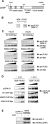
Figure 1
Figure 1. CHIP E3 ligase activity promotes the down-regulation of SGK-1
(A) Domain structure of WT and mutant CHIP. (B) SK-BR-3 cells were co-transfected with SGK-1–FLAG DNA (1.5 μg) and pcDNA3.1 (1.5 μg). At 36 h post-transfection, ALLN or vehicle (ethanol) were added overnight. The cells were lysed and subjected to immunoblotting with anti-FLAG antibodies to detect SGK-1. (C) The plasmids encoding SGK-1 (1.5 μg) and increasing amounts of WT, H260Q and K30A CHIP–Myc (0.1, 0.2, 0.38 and 0.75 μg) were expressed in SK-BR-3 cells. ALLN was added as above, and the cells were lysed and subjected to Western blot analysis with the indicated antibodies. (D) Increasing amounts of SGK-1–FLAG-encoding plasmids (0.5, 1.0 and 2.0 μg) were co-transfected with WT and mutant CHIP constructs into SK-BR-3 cells and SGK-1 expression was determined by Western blot analysis. (E) MDA-MB-231 cells stably expressing H260Q CHIP or the empty pcDNA3.1 vector were treated with ALLN or vehicle overnight and then examined by Western blotting for endogenous SGK-1 and ectopic CHIP expression. IB, immunoblot.
Figure 2
Figure 2. CHIP and Hsp/Hsc70 interact with SGK-1 in vivo
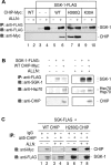
(A) SGK-1–FLAG was expressed in SK-BR-3 cells and immunoprecipitated followed by Western blotting in the presence or absence of WT and mutant CHIP–Myc expression. (B) SGK-1 was immunoprecipitated from SK-BR-3 cells stably expressing SGK-1–FLAG and transiently expressing WT CHIP–Myc. Immunoblotting was then performed to detect SGK-1 and Hsp/Hsc70 or CHIP using the appropriate antibody. (C) WT or H260Q CHIP was immunoprecipitated from SK-BR-3 cells transiently transfected with SGK-1–FLAG and CHIP constructs and subjected to Western blot analysis to detect SGK-1. IB, immunoblot.
Figure 3
Figure 3. CHIP is required for SGK-1 protein stability and ubiquitin modification
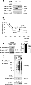
(A) CHIP+/+ and CHIP−/− mouse fibroblasts were treated overnight with ALLN or vehicle, lysed, and then examined by Western blotting for endogenous SGK-1, CHIP, Akt-1 and phospho-Akt-1 proteins. (B) Mouse fibroblasts were treated with 1 μg/ml of actinomycin D for indicated time periods. Total RNA was then isolated, and SGK-1 mRNA was quantified using quantitative real-time PCR. The ratio of SGK-1 to Actb mRNAs was calculated at each time point, and then expressed as a percentage of SGK-1/Actb ratio at 60 min. Data points represent the means for three independent experiments. S.E.M. and the calculated half-life are shown. (C) SK-BR-3 cells were stably transfected with CHIP siRNA and subjected to immunoblotting to examine endogenous SGK-1, CHIP and tubulin levels. The representative immunoblot from one of three independent experiments is shown. Quantification of the representative immunoblot shows CHIP protein levels relative to those of tubulin. (D) SK-BR-3 cells stably expressing SGK-1–FLAG were transfected with HA–ubiquitin plasmid and increasing amounts of WT CHIP plasmid. Immunoprecipitated SGK-1 was subjected to immunoblotting with anti-SGK-1 and anti-HA antibodies. CHIP expression was evaluated by Western blot analysis in the total lysate. The blot is representative of three independent experiments. IB, immunoblot.
Figure 4
Figure 4. SGK-1 and CHIP co-localize to the ER
(A) COS-7 cells were transfected with the plasmids encoding SGK-1–GFP, WT-CHIP–Myc or both (top, middle and bottom panels respectively) and the ER marker, pDsRed2-ER (which encodes calreticulin and an ER-retention signal fused to DsRed2). CHIP was detected using an anti-Myc antibody and a secondary anti-mouse antibody conjugated to Alexa Fluor® 488 (middle and bottom panels). (B) COS-7 cells were transfected with pDsRed2-ER vector (top and middle panels). Endogenous SGK-1 was detected with an anti-SGK-1 chicken antibody followed by an anti-chicken Alexa Fluor® 488-conjugated secondary antibody (top and bottom panels). Endogenous CHIP was detected with anti-CHIP rabbit antibody followed by anti-rabbit Alexa Fluor® 488-conjugated secondary antibody (middle panel) or anti-rabbit Alexa Fluor® 568-conjugated secondary antibody (bottom panel). Co-localization is shown in the merge column in yellow.
Figure 5
Figure 5. CHIP expression levels regulate SGK-1 in vitro kinase activity
(A) Following transient transfection of SK-BR-3 cells with the plasmids encoding SGK-1–FLAG and either CHIP–Myc or CHIP siRNA, SGK-1 was immunoprecipitated and used in an in vitro kinase assay as previously described [17]. Ratios of SGK-1 activity in the presence of CHIP–Myc or CHIP siRNA to SGK-1 activity alone are shown as a mean of six or four independent experiments respectively±S.E.M. *P<0.05 compared with SGK-1 activity alone. (B) Following the kinase assay, levels of immunoprecipitated SGK-1 were determined by Western blot analysis. In the total lysates, ectopic CHIP was detected with an anti-Myc antibody, while endogenous CHIP and tubulin were determined using an anti-CHIP and an anti-tubulin antibody respectively. IP, immunoprecipitation; IB, immunoblot.
Figure 6
Figure 6. SGK-1 and CHIP have both overlapping and independent anti-apoptotic roles
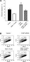
(A) SK-BR-3 cells stably expressing SGK-1–FLAG or empty vector were transiently transfected with CHIP siRNA DNA. At 48 h post UV-irradiation (100 J/m2), the percentage of apoptotic cells was determined using flow cytometric detection of cells staining for cleaved caspase-3 using the respective Alexa Fluor® 488-conjugated antibody. Results represent the means of the positively stained cell population±S.E.M. for three independent experiments. (B) Representative flow cytometric plots depict the percentage of cleaved caspase-3-positively stained cells in the experimental groups.
Figure 7
Figure 7. Proposed model of CHIP-mediated regulation of SGK-1 function
SGK-1 is induced as part of the stress response and is then available to be phosphorylated and activated downstream of PI3K. Upon either a direct or indirect interaction with CHIP at or near the ER, we hypothesize that SGK-1 becomes properly folded and fully active, thereby promoting cell survival. CHIP-mediated ubiquitin (Ub) modification of SGK-1, however, also allows for the subsequent rapid degradation and return to baseline of SGK-1 levels.

References

    1. Lang F., Cohen P. The regulation and physiological roles of serum and glucocorticoid-induced protein kinase. Science STKE 2001, RE17. 2001 - PubMed
    1. Hertweck M., Gobel C., Baumeister R. C. elegans SGK-1 is the critical component in the Akt/PKB kinase complex to control stress response and life span. Dev. Cell. 2004;6:577–588. - PubMed
    1. Lamitina S. T., Strange K. Transcriptional targets of DAF-16 insulin signaling pathway protect C. elegans from extreme hypertonic stress. Am. J. Physiol. Cell Physiol. 2005;288:C467–C474. - PubMed
    1. Mikosz C. A., Brickley D. R., Sharkey M. S., Moran T. W., Conzen S. D. Glucocorticoid receptor-mediated protection from apoptosis is associated with induction of the serine/threonine survival kinase gene, sgk-1. J. Biol. Chem. 2001;276:16649–16654. - PubMed
    1. Wu W., Chaudhuri S., Brickley D. R., Pang D., Karrison T., Conzen S. D. Microarray analysis reveals glucocorticoid-regulated survival genes that are associated with inhibition of apoptosis in breast epithelial cells. Cancer Res. 2004;64:1757–1764. - PubMed

Publication types

MeSH terms