. 2006 Oct 13;281(41):31021-30.
doi: 10.1074/jbc.M606278200. Epub 2006 Aug 7.

Binding of myotrophin/V-1 to actin-capping protein: implications for how capping protein binds to the filament barbed end

Affiliations

Binding of myotrophin/V-1 to actin-capping protein: implications for how capping protein binds to the filament barbed end

Nandini Bhattacharya et al. J Biol Chem. .

Abstract

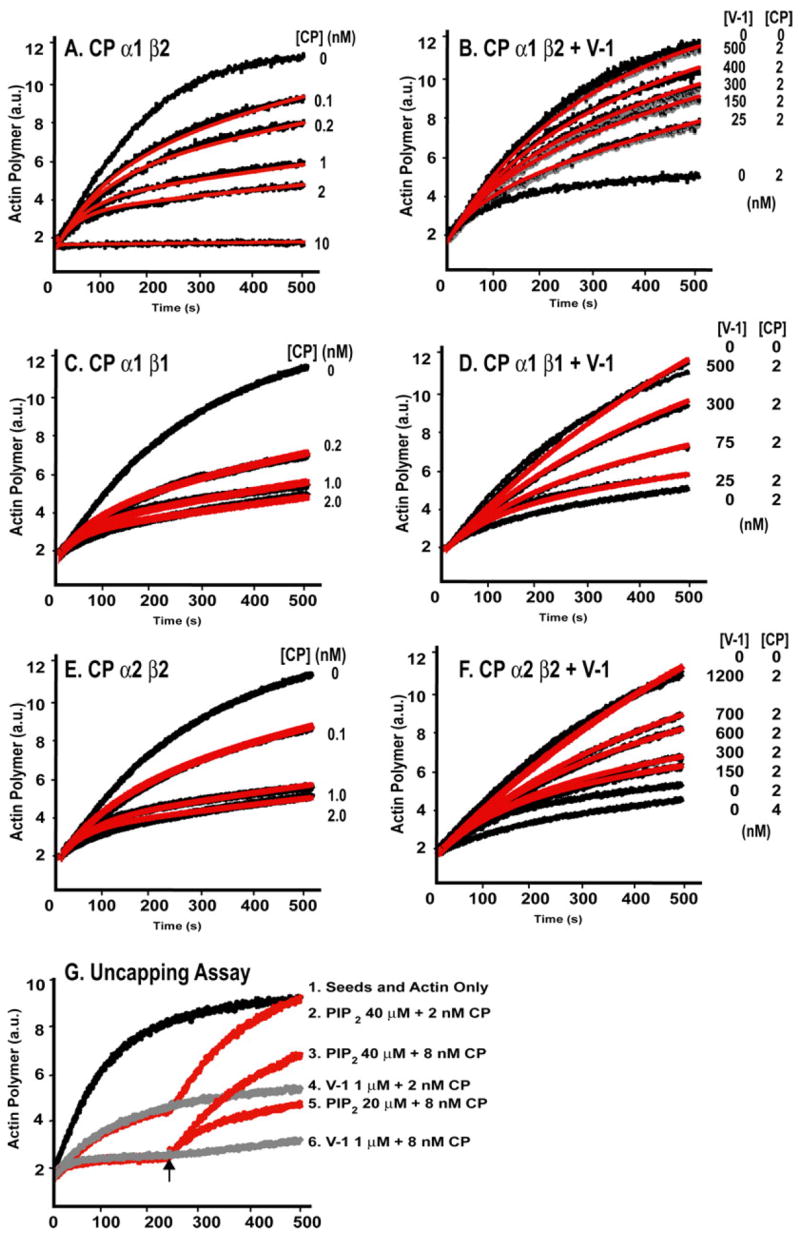
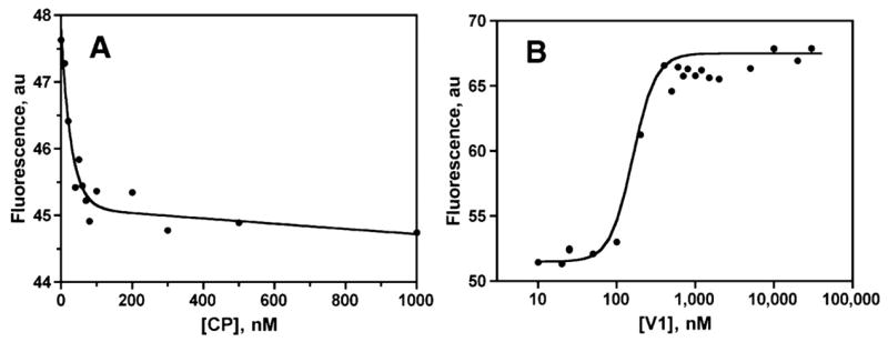
The heterodimeric actin-capping protein (CP) regulates actin assembly and cell motility by binding tightly to the barbed end of the actin filament. Here we demonstrate that myotrophin/V-1 binds directly to CP in a 1:1 molar ratio with a Kd of 10-50 nm. V-1 binding inhibited the ability of CP to cap the barbed ends of actin filaments. The actin-binding COOH-terminal region, the "tentacle," of the CP beta subunit was important for binding V-1, with lesser contributions from the alpha subunit COOH-terminal region and the body of the protein. V-1 appears to be unable to bind to CP that is on the barbed end, based on the observations that V-1 had no activity in an uncapping assay and that the V-1.CP complex had no capping activity. Two loops of V-1, which extend out from the alpha-helical backbone of this ankyrin repeat protein, were necessary for V-1 to bind CP. Parallel computational studies determined a bound conformation of the beta tentacle with V-1 that is consistent with these findings, and they offered insight into experimentally observed differences between the alpha1 and alpha2 isoforms as well as the mutant lacking the alpha tentacle. These results support and extend our "wobble" model for CP binding to the actin filament, in which the two COOH-terminal regions of CP bind independently to the actin filament, and bound CP is able to wobble when attached only via its mobile beta-subunit tentacle. This model is also supported by molecular dynamics simulations of CP reported here. The existence of the wobble state may be important for actin dynamics in cells.


Figures

FIGURE 1
FIGURE 1. Molecular dynamics analysis of CP
Range of dynamics of the α and β COOH termini (tentacles) based on a 20-ns molecular dynamics simulation.
FIGURE 2
FIGURE 2. Molecular dynamics analysis of the effect of truncation of the CP αCOOH terminus
A, regions on the α subunit that show a significant difference in dynamics upon deletion of the αtentacle are shown in blue, orange, and purple, and those colors correspond to the bars in C. The translucent pink region represents the surface where V-1 is predicted to contact the α subunit. B, residues that differ between the α1 and α2 isoforms are labeled and shown in space-filling mode. They overlap extensively with the pink contact surface and with the colored increased fluctuation residues of A. C and D, root mean square fluctuations of the α and β subunit residues during a 20-ns MD simulation. Wild-type CP is compared with a CP mutant lacking the α subunit COOH-terminal region. C, the α subunit shows three regions of significant change, indicated by colors that match the corresponding regions in B. D, the β subunit shows only one small difference, in an α-helical segment that underlies the α-subunit COOH-terminal region, colored green on the graph and in the inset.
FIGURE 3
FIGURE 3. Inhibition of CP by V-1 in actin polymerization seeded growth assays
Actin polymer concentration based on fluorescence (arbitrary units; a.u.) of pyrene-labeled actin (2 μM; 4% pyrene) is plotted versus time. Polymerization was nucleated with spectrin-actin seeds. A, C, and E, CP α1β2, α1β1, and α2β2, respectively, cap the barbed end of actin at indicated concentrations (black lines). Best fit curves from kinetic modeling with Reactions 1 and 2(see “Experimental Procedures”) are shown in red. B, D, and F, V-1 inhibits capping by CP α1β2, α1β1, and α2β2, respectively. Actin polymerization is shown versus time with CP and V-1 concentrations as indicated. Best fit curves from kinetic modeling with Reactions1–3 (see “Experimental Procedures”)are shown in red. G,V-1does not uncap barbed ends capped with CP. Actin polymerization versus time in the presence of 2 or 8 nM CP. After 250 s, PIP2 or GST-V-1 was added (arrow). PIP2 causes rapid uncapping, and GST-V-1 has no effect.
FIGURE 4
FIGURE 4. V-1 inhibition of CP in a steady-state assay
Pyrene-actin fluorescence, which is proportional to the concentration of filamentous actin, is plotted. A, increasing concentrations of CP α1β2, in the absence of V-1, causes the F-actin level to fall, because the critical concentration rises to that of the pointed end. B, increasing concentrations of V-1 added to 75 nM CP antagonize this inhibitory effect and promote actin polymerization.
FIGURE 5
FIGURE 5. Continuous variation analysis of the stoichiometry of V-1 interaction with CP, assayed by intrinsic tryptophan fluorescence
Fluorescence in arbitrary units (a.u.) is plotted against the concentrations of the proteins. Values from two independent experiments are shown.
FIGURE 6
FIGURE 6. V-1 inhibition of CP COOH-terminal truncation mutants in actin polymerization growth assays
Actin polymer concentration based on arbitrary units of pyrene fluorescence is plotted versus time. A, CPα1(Δ28) β1; B, CPα1β1(ΔC34). Conditions were as in Fig. 1, with the indicated concentrations of V-1 and mutant CP. Best fits are shown in red from modeling with Reactions 1–3 (see “Experimental Procedures”). C, V-1 interaction with the CPα1(Δ28) β1(Δ34) mutant. This CP mutant does not cap actin, but it competes with WT CP for binding to V-1. Concentrations of the proteins (in nM) were as indicated on the right. Best fits from kinetic modeling, shown in red, were determined with Reactions 1– 4 (see “Experimental Procedures”).
FIGURE 7
FIGURE 7. V-1 interaction with peptides corresponding to the COOH-terminal region of the β subunit, both β1 and β2 isoforms, assayed with actin polymerization growth assays (2 μM actin, 4% pyrene-labeled)
A, high concentrations of the 34-aa β2 peptide are able to cap. B, the addition of V-1 at concentrations up to 6 μM inhibits the capping activity, almost completely. C, the 28-aa β1 peptide caps actin at 5 μM, with partial inhibition by V-1 at concentrations up to 1.5 μM. D, the 34-aa β1 peptide partially caps actin at 1.5 μM, with partial inhibition by V-1 at concentrations up to 0.8 μM.
FIGURE 8
FIGURE 8. Effect of V-1 mutants on capping activity of CP
A, NMR solution structure of V-1 adapted from Yang et al. (11). The α helices are labeled as α 1–α8. The loops of the complete ankyrin repeats are labeled ANK1 and ANK2. Residues EGGR (arrow; ANK1) and KHHI (arrow; ANK2) were mutated to alanine. Arrows mark the start of the truncation mutants V-1 Δ1–20 (deletion of the first 20 amino acids) and V-1 Δ1–94 (deletion of the first 94 amino acids). B, alignment of V-1 sequences from various organisms, with some features from A labeled. A period indicates a residue identical to the one in the top line, mouse V-1. The mutated loop regions, boxed, have very similar sequences among vertebrates. The alignment was performed with ClustalW. For C–F, actin polymerization is plotted versus time for a seeded growth assay. 2 μM actin, 4% pyrene-labeled, was used with indicated concentrations of V-1 mutants and mouse CP α1β2. The best fit from kinetic modeling of Reactions 1–3 (see “Experimental Procedures”) is shown in red. C, effect of the V-1 ANK1 mutant, with a magnified view of the early time course of the reactions. D, effect of the V-1 ANK2 mutant, again with a magnified view of early time. E, effect of the V-1 Δ1–94 mutant. F, effect of the V-1 Δ1–20 mutant.
FIGURE 9
FIGURE 9. Model illustrating the interaction of V-1 and CP based on computational docking
A, the COOH-terminal tentacle of CP β1 (green) associates with V-1 (pink). B, magnified view of several residues important in the interaction, including the two loops of V-1.

Similar articles

Cited by

References

    1. Sen S, Kundu G, Mekhail N, Castel J, Misono K, Healy B. J Biol Chem. 1990;27:16635–16643. - PubMed
    1. Sil P, Misono K, Sen S. Circ Res. 1993;1:98–108. - PubMed
    1. Sarkar S, Leaman DW, Gupta S, Sil P, Young D, Morehead A, Mukherjee D, Ratliff N, Sun Y, Rayborn M, Hollyfield J, Sen S. J Biol Chem. 2004;19:20422–20434. - PubMed
    1. Pennica D, Shaw KJ, Luoh SM, Wood WI. Gene (Amst) 1995;158:305–306. - PubMed
    1. Taoka M, Isobe T, Okuyama T, Watanabe M, Kondo H, Yamakawa Y, Ozawa F, Hishinuma F, Kubota M, Minegishi A. J Biol Chem. 1994;269:9946–9951. - PubMed

Publication types

Substances