. 2006 Aug 15;103(33):12371-6.
doi: 10.1073/pnas.0605418103. Epub 2006 Aug 8.

Glycoproteomic probes for fluorescent imaging of fucosylated glycans in vivo

Affiliations

Glycoproteomic probes for fluorescent imaging of fucosylated glycans in vivo

Masaaki Sawa et al. Proc Natl Acad Sci U S A. .

Abstract

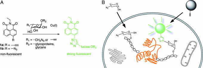
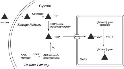
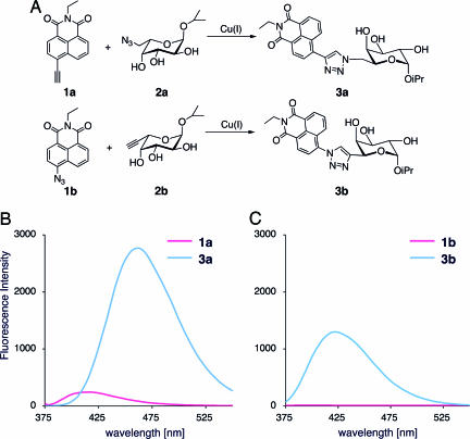
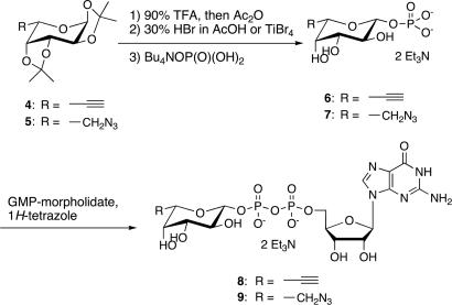
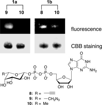
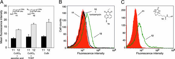
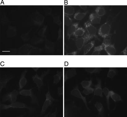
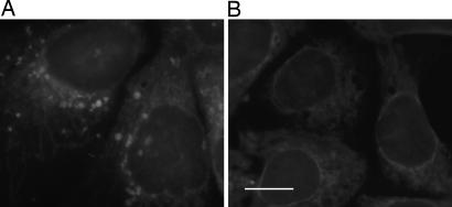
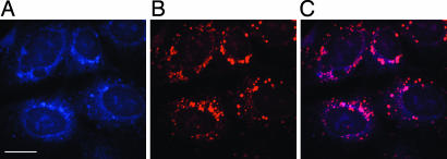
Glycomics is emerging as a new field for the biology of complex glycoproteins and glycoconjugates. The lack of versatile glycan-labeling methods has presented a major obstacle to visualizing at the cellular level and studying glycoconjugates. To address this issue, we developed a fluorescent labeling technique based on the Cu(I)-catalyzed [3 + 2] cycloaddition, or click chemistry, which allows rapid, versatile, and specific covalent labeling of cellular glycans bearing azide groups. The method entails generating a fluorescent probe from a nonfluorescent precursor, 4-ethynyl-N-ethyl-1,8-naphthalimide, by clicking the fluorescent trigger, the alkyne at the 4 position, with an azido-modified sugar. Using this click-activated fluorescent probe, we demonstrate incorporation of an azido-containing fucose analog into glycoproteins via the fucose salvage pathway. Distinct fluorescent signals were observed by flow cytometry when cells treated with 6-azidofucose were labeled with the click-activated fluorogenic probe or biotinylated alkyne. The intracellular localization of fucosylated glycoconjugates was visualized by using fluorescence microscopy. This technique will allow dynamic imaging of cellular fucosylation and facilitate studies of fucosylated glycoproteins and glycolipids.


Conflict of interest statement

Conflict of interest statement: No conflicts declared.

Figures

Fig. 1.
Fig. 1.
General strategy for glycan labeling. (A) Probe structures based on 1,8-naphthalimide include an azide or alkyne at a position of the ring that will allow a fluorogenic ligation with 6-modified fucose analogs. The fluorescence adduct is generated when probes are reacted with the azido/alkynyl group of fucosides via Cu(I)-catalyzed [3 + 2] cycloaddition. (B) Strategy for specific fluorescent labeling of fucosylated glycans in cells. Covalent modification of the target glycan with probes 1a or 1b results in production of fluorescently labeled glycoproteins bearing modified fucose (azidofucose shown).
Fig. 2.
Fig. 2.
Biosynthetic pathways for GDP-fucose. The de novo pathway transforms GDP-mannose into GDP-fucose via two enzymes, GDP-mannose 4,6-dehydratase (GMD) and GDP-keto-6-deoxymannose 3,5-epimerase/4-reductase (FX protein). The salvage pathway utilizes free fucose in the cytosol to create GDP-fucose by the action of fucose kinase and GDP-fucose pyrophosphorylase. The resulting GDP-fucose is used by fucosyltransferases (FucTs) in the Golgi apparatus to catalyze transfer onto glycoconjugates.
Fig. 3.
Fig. 3.
The “click” reaction of probes 1a and 1b with fucose derivatives results in highly fluorescent adducts (A) and fluorescence spectra of compounds 1a and 1b and their click products 3a and 3b (B and C).
Scheme 1.
Scheme 1.
Synthesis of GDP-fucose analogs.
Fig. 4.
Fig. 4.
Visualization of AGP after FucT transfer of modified GDP-fucose and labeling reaction with fluorogenic probes 1a and 1b. Treated protein was separated by SDS/PAGE and visualized by UV light (Top) and Coomassie blue staining (CBB) (Middle). (Bottom) Modified GDP-fucose.
Fig. 5.
Fig. 5.
Analysis of fucosylated glycoproteins on the cell surface of Jurkat cells by flow cytometry. (A) Comparison of labeling efficiency by using various copper sources including CuSO2/ascorbic acid, CuSO4/TCEP, or CuBr. Data points represent the average of triplicate experiments. (B) Treatment with tunicamycin suppressed cell surface fluorescence (cells cultured with natural fucose in red, azidofucose in green, or azidofucose in the presence of tunicamycin in black). (C) Labeling with biotinylated alkyne 13 was performed with UltraAvidin-Fluorescein, then analyzed by flow cytometry (cells treated with fucose in red or azidofucose in green).
Fig. 6.
Fig. 6.
Fluorescent image of cells labeled with probe 1a. The labeling reaction was performed in the presence of Cu(I) after the treatment with natural fucose (A) or azidofucose (B), or in the absence of Cu(I) after the treatment with natural fucose (C) or azidofucose (D). (Scale bar: 20 μm.)
Fig. 7.
Fig. 7.
Specificity of the Cu(I)-catalyzed cycloaddition for azidofucose. Azidofucose-supplemented cells were fixed and treated directly with probe (A) or were reduced with Tris(3-sulfonatophenyl)phosphine and then subjected to labeling reaction (B). (Scale bar: 20 μm.)
Fig. 8.
Fig. 8.
Imaging intracellular fucosylation by double staining with the probe 1a and WGA lectin. Azidofucose treated cells were fixed and labeled with probe 1a and then further treated with WGA lectin conjugated with Alexa Fluor 594, and the cells were imaged with confocal fluorescence microscope by using appropriate filter sets. (A) Blue fluorescence, labeled with probe 1a. (B) Red fluorescence, stained with Alexa Fluor 594-conjugated WGA lectin as a Golgi marker. (C) Purple color, overlap in blue and red signals. (Scale bar: 20 μm.)

Similar articles

Cited by

References

    1. Apweiler R., Hermjakob H., Sharon N. Biochim. Biophys. Acta. 1999;1473:4–8. - PubMed
    1. Becker D. J., Lowe J. B. Glycobiology. 2003;13:41R–53R. - PubMed
    1. Staudacher E. Trends Glycosci. Glycotechnol. 1996;8:391–408.
    1. Sears P., Wong C.-H. Cell. Mol. Life Sci. 1998;54:223–252. - PMC - PubMed
    1. Haltiwanger R. S., Lowe J. B. Annu. Rev. Biochem. 2004;73:491–537. - PubMed

Publication types