. 2006 Aug 15;103(33):12592-7.
doi: 10.1073/pnas.0602032103. Epub 2006 Aug 8.

Viral tracing identifies distributed columnar organization in the olfactory bulb

Affiliations

Viral tracing identifies distributed columnar organization in the olfactory bulb

David C Willhite et al. Proc Natl Acad Sci U S A. .

Abstract

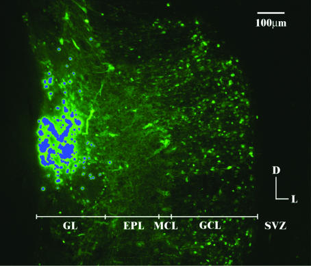
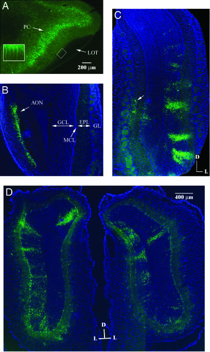
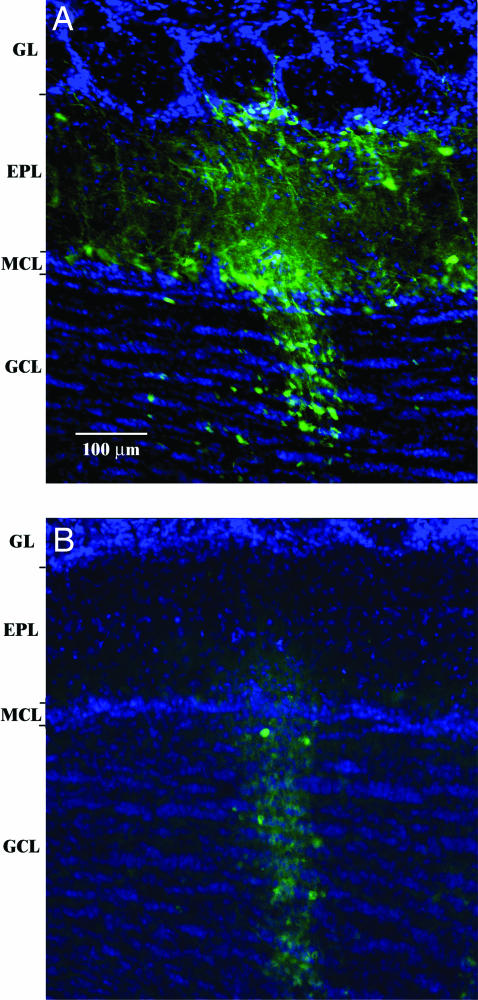
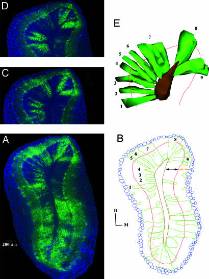
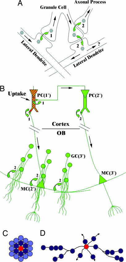
Olfactory sensory neurons converge onto glomeruli in the olfactory bulb (OB) to form modular information processing units. Similar input modules are organized in translaminar columns for other sensory modalities. It has been less clear in the OB whether the initial modular organization relates to a columnar structure in the deeper layers involved in local circuit processing. To probe synaptic connectivity in the OB, we injected a retrograde-specific strain of the pseudorabies virus into the rat OB and piriform cortex. The viral-staining patterns revealed a striking columnar organization that extended across all layers of the OB from the glomeruli to the deep granule cell layer. We hypothesize that the columns represent an extension of the glomerular unit. Specific patterning was observed, suggesting selective, rather than distance-dependent, center-surround connectivity. The results provide a previously undescribed basis for interpreting the synaptic connections between mitral and granule cells within the context of a columnar organization in the OB and have implications for olfactory coding and network organization.


Conflict of interest statement

Conflict of interest statement: No conflicts declared.

Figures

Fig. 1.
Fig. 1.
An OB injection site. Coronal section of the rat brain 3 days after PRV injection into the anterior dorsomedial OB viewed at ×100 magnification. Virus-infected cells are visible in green (GFP), green fluorescent beads were included as a marker of the injection site, and they are easily distinguished from viral staining in practice by intensity and punctate appearance. Beads are false colored blue for clarity. GL, glomerular layer; EPL, external plexiform layer; MCL, mitral cell layer; SVZ, subventricular zone. Orientation bar indicates dorsal and lateral.
Fig. 2.
Fig. 2.
PRV staining patterns after OB injections. A-C) Coronal sections of the rat brain 3 days after PRV injection into the anterior dorsolateral OB at ×40 magnification. Virus-infected cells are stained green (GFP and α-GFP), and all cell nuclei in B and C are blue (DAPI). Scale bar in A applies to AC. (A) The ipsilateral piriform cortex (PC) is prominently labeled in this section ≈3.7 mm anterior to Bregma, whereas the LOT is not. Note, however, several processes penetrating the LOT (Inset, ×200 magnification of boxed region). (B) A section showing the contralateral bulb ≈6.5 mm anterior to Bregma. Only the anterior olfactory nucleus (AON) contains PRV. MCL, mitral cell layer; EPL, external plexiform layer; GL, glomerular layer. (C) A section of the ipsilateral bulb posterior to the injection site. The majority of PRV-stained cells appear in the injection-side half of the OB in radial patterns. The arrow shows a labeled tufted cell in the medial side, possibly an intrabulbar projection neuron identified by Belluscio et al. (50). (D) Coronal section of the rat OB 3 days after PRV injections into the anterior ventrolateral (Left) and anterior ventromedial (Right) OBs of the same animal (overlay of two fields at ×40 magnification). As with the anterior dorsal injections, the majority of columns appear in the ipsilateral half of the OB.
Fig. 3.
Fig. 3.
Columns with and without associated mitral/tufted cells. Coronal sections of the rat OB 3 days after PRV injection into the dorsomedial OB (two fields overlain, colored as in Fig. 2). (A) A column with associated mitral and tufted cells. Mitral and tufted cells are labeled without granule cell columns to both sides of the full column. (B) A granule cell-only column with no mitral or tufted cells stained.
Fig. 4.
Fig. 4.
OB columns from anterior piriform cortex PRV injection. (A) Coronal section of the rat OB 3 days after injection of PRV into the anterior piriform cortex, colored as in Fig. 2. (B) Schematic of the OB organization of A. Blue, glomeruli; red, mitral cell layer (MCL); brown, subventricular zone; green, virus-labeled columns. Black arrows indicate the GCL; orange arrows the external plexiform layer. Dashed green lines indicate areas that may be interpreted as further division of a larger column. Columns 5 and 6 extend from the glomerular layer to the deepest area of the GCL nearly completely within the confines of the 60-μm section shown. Columns 1–4 also extend from the deep GCL to single glomeruli residing in other sections. (C and D) Adjacent sections anterior to A, showing the progression of columns 2–4. (E) A 3D reconstruction of the selected columns through 11 sections, 5 anterior and 5 posterior to A. The red line represents the MCL in A for reference.
Fig. 5.
Fig. 5.
Models for PRV transfer and staining patterns in the OB. (A) A diagram of the retrograde synaptic transfer of PRV (hexagrams) in OB neurons. Virus comes from the soma of a first-order infected mitral cell to the left and travels down the lateral dendrite to a granule cell, crossing the first synapse (1 in diagram). Virus then replicates within the granule cell and passes to axonal processes, including mitral cell axon collaterals and centrifugal fibers (2 at top). Because of the reciprocal nature of the GC synapse, virus also may pass to another secondary dendrite (2 at bottom), although it is not known whether productive infection can occur through this transmission route. (B) A diagram showing an interpretation of the viral-staining patterns from piriform cortex injection. The virus is taken up at the injection site by pyramidal cells (PC) in the cortex (red and green cell, top left; 1′, 2′ and 3′ refer to first-, second-, and third-order neurons, respectively). After 24 h, the virus replicates and crosses the synapses to axons from the cortical cells dendrites (1 in diagram). The virus travels in a retrograde manner to other pyramidal cells in the cortex through the cortico-cortical connections and to mitral cells (MC) in the OB through the lateral olfactory tract (tufted and periglomerular cells are omitted for simplification). Twenty-four hours later, virus is visible by GFP fluorescence in the second-order neurons, and the second synapses are crossed (2 in diagram). Another 24 h shows fluorescence in the granule cells (GC) that represent third-order neurons (Left) and lone mitral cells that are also third order neurons infection from second order pyramidal cells (Right). Note that the rightmost granule cell population is labeled from infection by the lateral dendrite from the second-order mitral cell on the left, not the third order mitral cell on the right. (C and D) Distance-dependent center-surround vs. selective inhibition. Circles represent a top view of glomerular units. (C) In a distance-dependent center-surround inhibition model, the activated glomerulus (red) inhibits surrounding glomeruli in a distance-dependent manner. Inhibition is shown by the intensity of blue. (D) In a selective inhibition model, the activated glomerulus inhibits selectively connected glomeruli through the synapses on the mitral and tufted cell secondary dendrites at the granule cell level. Such connections would extend in all directions from the activated glomerulus (arrows). This model is consistent with a recent computational models of odor coding (36, 51).

Similar articles

Cited by

References

    1. Lorente de No R. In: Physiology of the Nervous System, Fulton J. F., editor. London: Oxford Univ. Press; 1938. pp. 291–339.
    1. Mountcastle V. B. J. Neurophysiol. 1957;20:408–434. - PubMed
    1. Hubel D. H., Wiesel T. N. J. Physiol. 1968;195:215–243. - PMC - PubMed
    1. Hubel D. H., Wiesel T. N. J. Physiol. 1962;160:106–154. - PMC - PubMed
    1. Imig T. J., Adrian H. O. Brain Res. 1977;138:241–257. - PubMed

Publication types

LinkOut - more resources