. 2006 Aug;5(8):1215-28.
doi: 10.1128/EC.00037-06.

Analysis of mitogen-activated protein kinase signaling specificity in response to hyperosmotic stress: use of an analog-sensitive HOG1 allele

Affiliations

Analysis of mitogen-activated protein kinase signaling specificity in response to hyperosmotic stress: use of an analog-sensitive HOG1 allele

Patrick J Westfall et al. Eukaryot Cell. 2006 Aug.

Abstract

When confronted with a marked increase in external osmolarity, budding yeast (Saccharomyces cerevisiae) cells utilize a conserved mitogen-activated protein kinase (MAPK) signaling cascade (the high-osmolarity glycerol or HOG pathway) to elicit cellular responses necessary to permit continued growth. One input that stimulates the HOG pathway requires the integral membrane protein and putative osmosensor Sho1, which recruits and enables activation of the MAPK kinase kinase Ste11. In mutants that lack the downstream MAPK kinase (pbs2Delta) or the MAPK (hog1Delta) of the HOG pathway, Ste11 activated by hyperosmotic stress is able to inappropriately stimulate the pheromone response pathway. This loss of signaling specificity is known as cross talk. To determine whether it is the Hog1 polypeptide per se or its kinase activity that is necessary to prevent cross talk, we constructed a fully functional analog-sensitive allele of HOG1 to permit acute inhibition of this enzyme without other detectable perturbations of the cell. We found that the catalytic activity of Hog1 is required continuously to prevent cross talk between the HOG pathway and both the pheromone response and invasive growth pathways. Moreover, contrary to previous reports, we found that the kinase activity of Hog1 is necessary for its stress-induced nuclear import. Finally, our results demonstrate a role for active Hog1 in maintaining signaling specificity under conditions of persistently high external osmolarity.


Figures
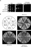
FIG. 1.
FIG. 1.
Phenotypic characterization of the hog1-as allele. (A) Tenfold serial dilutions of the indicated strains [HOG1, hog1Δ, hog1-as, hog1(K52R), and hog1(D144A)] were spotted onto plates containing YPD (left), YPD plus 1 M sorbitol (middle), or YPD plus 1 M sorbitol and 12 μM 1-NM-PP1 (right) and incubated at 30°C for 2 days. (B) The same HOG1, hog1Δ, or hog1-as mutant cells as in panel A were transformed with empty URA3-marked vector YEp352-GAL (upper half) or the same vector expressing Ssk2ΔN (lower half). Representative transformants were streaked on selective medium (synthetic complete medium without uracil) containing glucose (upper right), galactose (lower left), or galactose plus 12 μM 1-NM-PP1 (lower right) and incubated at 30°C for 2 days.
FIG. 2.
FIG. 2.
Biochemical characterization of the hog1-as allele. (A) HOG1, hog1Δ, and hog1-as mutant cells carrying an integrated copy of (HA)3 epitope-tagged Rck2 were grown to mid-exponential phase in YPD, pretreated in the absence (−) or presence (+) of 12 μM 1-NM-PP1 for 10 min, and then not exposed (−) or exposed (+) to 1 M sorbitol, as indicated. After 10 min, the cells were lysed as described in Materials and Methods and samples of the resulting extracts were analyzed by SDS-PAGE and immunoblotting with an anti-HA epitope antibody. 1, unmodified Rck2; 2 and 3, hyperphosphorylated Rck2. (B) The same strains as in Fig. 1A, each carrying an integrated copy of the FUS1prom-lacZ reporter gene, were grown to mid-exponential phase and then resuspended in YPD (lanes 1), YPD plus 1 M sorbitol (lanes 2), or YPD plus 1 M sorbitol and 12 μM 1-NM-PP1 (lanes 3), as indicated. After 5 h to allow for gene expression and enzyme synthesis, samples were assayed for β-galactosidase content as described in Materials and Methods.
FIG. 3.
FIG. 3.
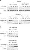
Kinetics of Hog1 phosphorylation in response to hyperosmotic stress. Cells expressing C-terminally GFP-tagged derivatives of wild-type Hog1 (A), Hog1-as (B), Hog1(K52R) (C), or Hog1(D144A) (D) were grown to mid-exponential phase in YPD and then exposed to 1 M sorbitol or 1 M sorbitol and 12 μM 1-NM-PP1, as indicated. At the times shown, samples of each culture were withdrawn and frozen immediately in liquid N2. For subsequent analysis, the samples were thawed on ice, lysed, resolved by SDS-PAGE, and subjected to immunoblotting with an anti-phospho-p38 antibody to detect active (dually phosphorylated) Hog1 (upper part of each panel) and an anti-GFP antibody to detect total Hog1 (bottom part of each panel).
FIG. 4.
FIG. 4.
Hog1 catalytic activity is required for its hyperosmotic stress-induced nuclear import. Cells expressing the same GFP-tagged versions of wild-type and mutant Hog1 as in Fig. 3 were grown to mid-exponential phase, prestained for 30 min at 30°C with 2 μM (final concentration) DAPI to visualize DNA, and then resuspended in YPD (left) or YPD plus 1 M sorbitol (right). After 10 min at 30°C, the cells were examined under an epifluorescence microscope. Arrows indicate the positions of representative nuclei.
FIG. 5.
FIG. 5.
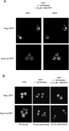
Analog inhibition blocks nuclear entry and does not stimulate nuclear exit. (A) Cells expressing C-terminally GFP-tagged wild-type Hog1 or Hog1-as were grown to mid-exponential phase in YPD, prestained with DAPI as described in the legend to Fig. 4, resuspended in YPD plus 12 μM 1-NM-PP1 for 10 min, and then exposed to 1 M (final concentration) sorbitol. After 10 min, the cells were examined under an epifluorescence microscope. (B) The same cells as in panel A were resuspended in YPD (left) or YPD plus 1 M sorbitol. After 10 min, samples of each were viewed with an epifluorescence microscope to confirm that hyperosmotic-stress-induced nuclear translocation had occurred in the sample exposed to 1 M sorbitol (middle). A portion of the sample that received 1 M sorbitol was then adjusted to 12 μM (final concentration) 1-NM-PP1. After 2 min and various times thereafter (up to 30 min), the cells that were exposed to 1 M sorbitol and 12 μM 1-NM-PP1 were examined again (right).
FIG. 6.
FIG. 6.
Nmd5 is required for nuclear import of Hog1 but does not itself enter the nucleus. (A) NMD5+ (top) and nmd5Δ mutant (bottom) cells expressing C-terminally GFP-tagged wild-type Hog1 were grown to mid-exponential phase in YPD and resuspended in YPD (left) or YPD plus 1 M sorbitol (right). After 10 min, the cells were viewed under a confocal fluorescence microscope. Representative 0.2-μm optical z sections are shown. (B) Cells coexpressing C-terminally GFP-tagged Hog1-as and fully functional C-terminally mCherry-tagged Nmd5 were grown to mid-exponential phase in YPD and resuspended in YPD (left), in YPD plus 1 M sorbitol (middle), or in YPD plus 1 M sorbitol and 12 μM 1-NM-PP1 (right). After 10 min, the cells were examined with the appropriate cutoff filters to view GFP (top) or mCherry (middle). The merged GFP and mCherry images are also shown (bottom).
FIG. 7.
FIG. 7.
Nmd5 and kinase activity are required for passage of Hog1 through the NPC. (A) An nmd5Δ nup133Δ double mutant coexpressing C-terminally GFP-tagged wild-type Hog1 and C-terminally mCherry-tagged Nup1 was grown to mid-exponential phase in YPD and resuspended in YPD plus 1 M sorbitol. After 10 min, the cells were examined with the appropriate cutoff filters to view GFP (left) or mCherry (middle). The merged GFP and mCherry images are also shown (right). (B) Cells expressing C-terminally GFP-tagged Hog1-as and C-terminally mCherry-tagged Nup1 were grown to mid-exponential phase in YPD and resuspended in YPD plus 1 M sorbitol and 12 μM 1-NM-PP1. After 10 min, the cells were examined with the appropriate cutoff filters to view GFP (left) or mCherry (middle). The merged GFP and mCherry images are also shown (right). In both the top and bottom parts, representative 0.2-μm optical z sections are shown.
FIG. 8.
FIG. 8.
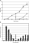
Persistent Hog1 function is required to prevent cross talk and maintain the adapted state. (A) Cells expressing Hog1-as were grown in YPD and, at time zero, resuspended in YPD plus 1 M sorbitol in the absence (filled circles) or presence (filled squares) of 12 μM 1-NM-PP1. After 3 h (arrow), 12 μM (final concentration) 1-NM-PP1 was added to a portion of the culture that was exposed to YPD plus 1 M sorbitol only (filled triangles). At the indicated times, samples were withdrawn and the level of expression of the FUS1prom-lacZ reporter was measured as described in the legend to Fig. 2B. (B) Cells expressing a C-terminally GFP-tagged version of wild-type Hog1 (gray bars) or Hog1-as (black bars) were grown in YPD and, at time zero, resuspended in YPD plus 1 M sorbitol. After 90 min (arrow), 12 μM (final concentration) 1-NM-PP1 was added to each culture. Samples were withdrawn at the indicated times, and the ratio of phospho-Hog1 to total Hog1 was determined as described in the legend to Fig. 3.
FIG. 9.
FIG. 9.
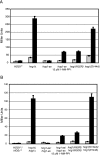
Hog1 catalytic activity is required to prevent cross talk with the filamentous-growth-signaling pathway. Isogenic TEC1+ haploid (A) and MATa/MATα diploid (B) derivatives of strain Σ1278b carrying an integrated TEC1prom-lacZ reporter and either wild-type HOG1 or the four mutant alleles described in the legend to Fig. 1A, as indicated, were grown to mid-exponential phase in YPD and then resuspended in YPD (gray bars) or YPD plus 1 M sorbitol (black bars). A portion of the hog1-as mutant cells was treated in the same way but also in the presence of 12 μM 1-NM-PP1, as shown. After 5 h, samples were assayed for β-galactosidase content as described in the legend to Fig. 2B.

Similar articles

Cited by

References

    1. Albertyn, J., S. Hohmann, J. M. Thevelein, and B. A. Prior. 1994. GPD1, which encodes glycerol-3-phosphate dehydrogenase, is essential for growth under osmotic stress in Saccharomyces cerevisiae, and its expression is regulated by the high-osmolarity glycerol response pathway. Mol. Cell. Biol. 14:4135-4144. - PMC - PubMed
    1. Bao, M. Z., M. A. Schwartz, G. T. Cantin, J. R. Yates III, and H. D. Madhani. 2004. Pheromone-dependent destruction of the Tec1 transcription factor is required for MAP kinase signaling specificity in yeast. Cell 119:991-1000. - PubMed
    1. Bardwell, L., J. G. Cook, E. C. Chang, B. R. Cairns, and J. Thorner. 1996. Signaling in the yeast pheromone response pathway: specific and high-affinity interaction of the mitogen-activated protein (MAP) kinases Kss1 and Fus3 with the upstream MAP kinase kinase Ste7. Mol. Cell. Biol. 16:3637-3650. - PMC - PubMed
    1. Benjamin, K. R., C. Zhang, K. M. Shokat, and I. Herskowitz. 2003. Control of landmark events in meiosis by the CDK Cdc28 and the meiosis-specific kinase Ime2. Genes Dev. 117:1524-1539. - PMC - PubMed
    1. Benton, B. M., W. K. Eng, J. J. Dunn, F. W. Studier, R. Sternglanz, and P. A. Fisher. 1990. Signal-mediated import of bacteriophage T7 RNA polymerase into the Saccharomyces cerevisiae nucleus and specific transcription of target genes. Mol. Cell. Biol. 10:353-360. - PMC - PubMed

Publication types

MeSH terms

Substances