

Morphological analysis of the proximal femur using quantitative computed tomography
- PMID: 16896872
- PMCID: PMC2267581
- DOI: 10.1007/s00264-006-0182-z
Morphological analysis of the proximal femur using quantitative computed tomography
Abstract
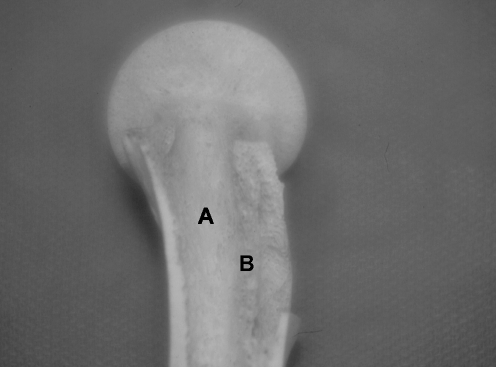
The anatomy of the proximal femur was studied in 35 specimens using quantitative computed tomography (QCT) and compared with anatomical sections studied by plane radiography and gross dissection. We found the primary supporting structure of the femoral head to be the primary compressive strut, which is a dense column of trabecular bone projecting from the pressure buttress of the medial femoral neck to the epiphyseal scar. Trabecular bone mushroomed from the epiphyseal scar and terminated at right angles to the cortex of the femoral head. We believe the primary compressive strut is the predominant load-bearing structure connecting the femoral head to the femoral neck, as many specimens lacked continuity of the head cortex to the femoral neck. Based on the CT number, the primary compressive strut had similar bone density to cortical structures such as the lesser trochanter, calcar femorale and posterior lateral femoral cortex. Ward's triangle lacked structural integrity in many cases, and we doubt the significance of tensile trabculae for sharing load. Surgical techniques such as femoral fracture fixation, resurfacing hip arthroplasty and allograft transplantation may benefit from this knowledge.
L’anatomie de l’extrémité supérieure du fémur a été étudiée chez 35 sujets à partir d’études de type scanner et comparée avec des sections anatomiques, des radiographies et des dissections. Nous avons étudié les différentes structures qui supporte la tête fémorale. Les coupes scanner montrent que la densité osseuse de ces structures est identique à celle du trochanter et de la corticale de la face postérieure du fémur. Les techniques chirurgicales concernant l’extrémité supérieure du fémur, fixation de fractures, resurfaçage, arthroplastie de resurfaçage et al. logreffes doivent bénéficier de ce travail anatomique et radiologique.
Figures


References
-
- {'text': '', 'ref_index': 1, 'ids': [{'type': 'DOI', 'value': '10.1359/jbmr.2000.15.12.2297', 'is_inner': False, 'url': 'https://doi.org/10.1359/jbmr.2000.15.12.2297'}, {'type': 'PubMed', 'value': '11127194', 'is_inner': True, 'url': 'https://pubmed.ncbi.nlm.nih.gov/11127194/'}]}
- Beck TJ, Looker AC, Ruff CB, Sievanen H, Wahner HW (2000) Structural trends in the aging femoral neck and proximal shaft: analysis ot the Third National Health and Nutrition Examination Survey dual-energy X-ray absorptiometry data. J Bone Miner Res 15:2297–2304 - PubMed
-
- {'text': '', 'ref_index': 1, 'ids': [{'type': 'PubMed', 'value': '2618148', 'is_inner': True, 'url': 'https://pubmed.ncbi.nlm.nih.gov/2618148/'}]}
- Bergmann G, Rohlmann A, Graichen F (1989) Invivo messung der Hüftgelenksbelastung: Teil: Krankengymnastik. Z Orthop 127:672–679 - PubMed
-
- {'text': '', 'ref_index': 1, 'ids': [{'type': 'PubMed', 'value': '7446022', 'is_inner': True, 'url': 'https://pubmed.ncbi.nlm.nih.gov/7446022/'}]}
- Brown TD, Ferguson AB (1980) Mechanical property distributions in the cancellous bone of the human proximal femur. Acta Scand Ortho 51:429–437 - PubMed
-
- {'text': '', 'ref_index': 1, 'ids': [{'type': 'PubMed', 'value': '8570492', 'is_inner': True, 'url': 'https://pubmed.ncbi.nlm.nih.gov/8570492/'}]}
- Chandler HP (1995) Structural grafting of the acetabulum. Orthopaedics 18:863–864 - PubMed
-
- {'text': '', 'ref_index': 1, 'ids': [{'type': 'DOI', 'value': '10.1016/0021-9290(89)90091-2', 'is_inner': False, 'url': 'https://doi.org/10.1016/0021-9290(89)90091-2'}, {'type': 'PubMed', 'value': '2722894', 'is_inner': True, 'url': 'https://pubmed.ncbi.nlm.nih.gov/2722894/'}]}
- Carter DR, Orr TE, Fyhrie DP (1989) Relationships between loading history and femoral cancellous bone architecture. J Biomech 22:231–244 - PubMed
MeSH terms
LinkOut - more resources
Full Text Sources
