

Review
doi: 10.1007/s00776-006-1037-6.
Premalignant conditions of bone
Affiliations
- PMID: 16897210
- PMCID: PMC2780648
- DOI: 10.1007/s00776-006-1037-6
Item in Clipboard
Review
Premalignant conditions of bone
J Orthop Sci.
2006 Jul.
No abstract available
Figures
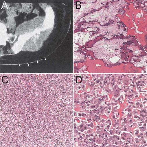
Osteosarcoma arising in Paget’s disease. A Left ileum with expansile mass in a patient with Paget’s disease. B Area of Paget’s disease uninvolved by sarcoma. C, D Osteo- genic sarcoma corresponding to iliac lesion with abundant hyperchromatic, pleomorphic cells and lace-like osteoid
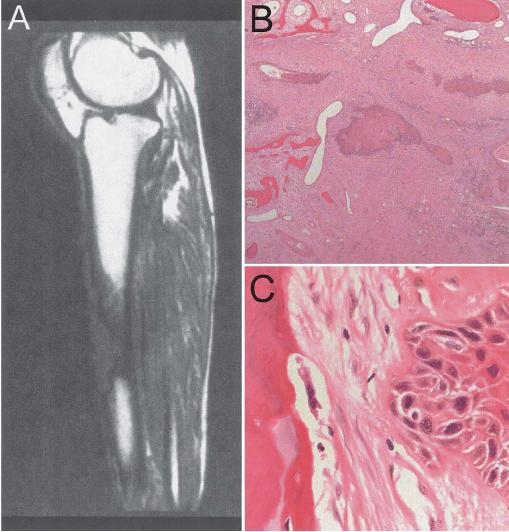
Squamous cell carcinoma arising in chronic osteomyelitis with a fistula tract. A Large, destructive lesion of the femur underneath a fistula tract noted on T1-weighted magnetic resonance imaging. B, C Squamous cell carcinoma shows diagnostic foci of keratinization and intercytoplasmic bridges
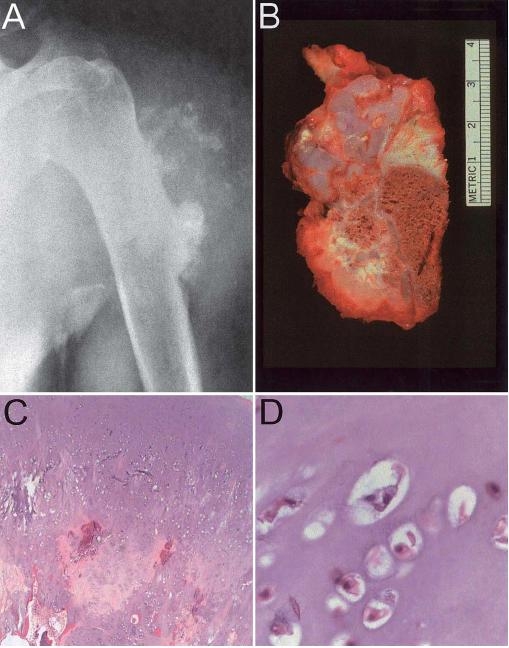
Chondrosarcoma arising in an osteochondroma. A Large, ill-defined soft tissue mass with ring-like calcifications arising at the site of prior osteochondroma in the humerus. B Gross pathology of chondrosarcoma arising from osteochondroma of the sacrum demonstrates irregular mineralization and cystic degeneration. C Loss of columnar architecture and thick cartilage cap (edge of cap is visible at top right) are common. D In a grade I chondrosarcoma cytology is typically bland
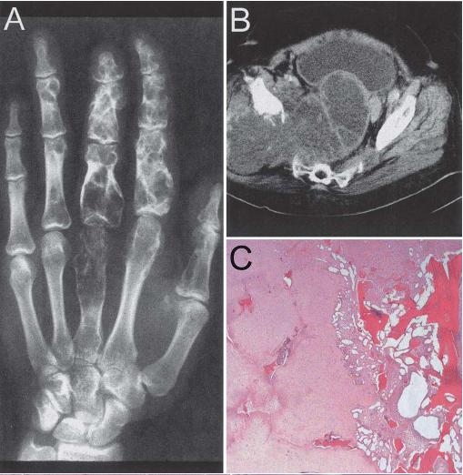
Chondrosarcoma arising in the setting of multiple enchondromas. A Plain radiograph from an Ollier disease patient demonstrating multiple enchondromas of the digits. B Computed tomography scan from an Ollier disease patient showing a large, destructive mass of the ileum. C Permeation of existing trabecular bone is diagnostic of chondrosarcoma
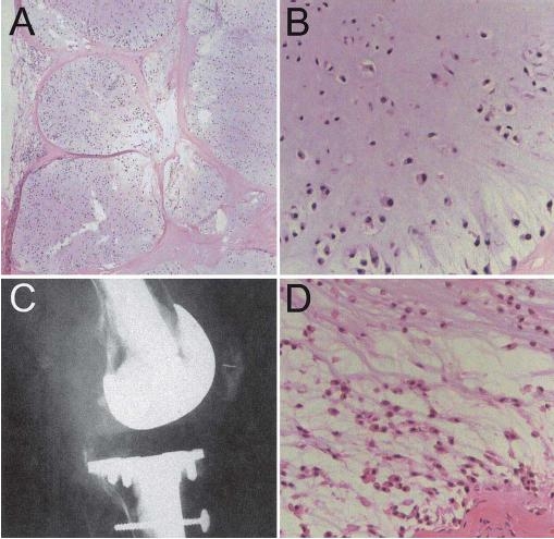
Chondrosarcoma in synovial chondromatosis. A The benign cartilage of synovial chondromatosis shows a lobular growth pattern at low power. B Synovial chondromatosis often demonstrates chondrocyte clustering, nuclear hyperchromasia, and binucleation and should not be mistaken for chondrosarcoma. C Plain radiograph of a patient with synovial chondromatosis and knee hemiarthroplasty with a new ill-defined soft tissue density adjacent to the knee joint. D Loss of clustering and spindling of chondrocytes at the periphery of a lobule are suggestive of chondrosarcoma in this setting
References
-
- Greditzer HG, 3rd, McLeod RA, Unni KK, Beabout JW. Bone sarcomas in Paget disease. Radiology. 1983;146:327–33. - PubMed
-
- McKenna RJ, Schwinn CP, Soong KY, Higinbotham NL. Sarcomate of the osteogenic series (osteosarcoma, fibrosarcoma, chondrosarcoma, parosteal osteogenic sarcoma, and sarcomata arising in abnormal bone): an analysis of 552 cases. J Bone Joint Surg. 1966;48:1–26.
Publication types
MeSH terms
LinkOut - more resources
Full Text Sources
Medical
