. 2006 Oct;99(1):206-17.
doi: 10.1111/j.1471-4159.2006.04096.x. Epub 2006 Aug 8.

Developmental and cell-selective variations in N-methyl-D-aspartate receptor degradation by calpain

Affiliations

Developmental and cell-selective variations in N-methyl-D-aspartate receptor degradation by calpain

Yi Na Dong et al. J Neurochem. 2006 Oct.

Abstract

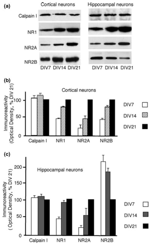
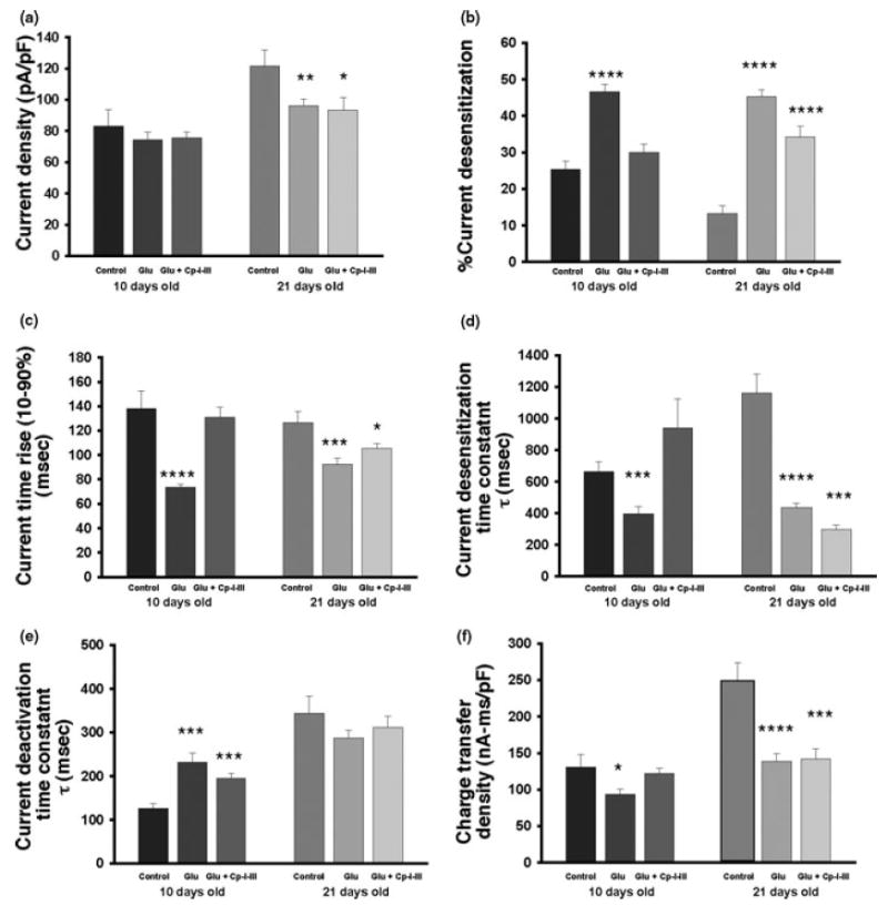
NMDA receptors play critical roles in synaptic modulation and neurological disorders. In this study, we investigated the developmental changes in NR2 cleavage by NMDA receptor-activated calpain in cultured cortical and hippocampal neurons. Calpain activity increased with development, associated with increased expression of NMDA receptors but not of calpain I. The activation of calpain in immature and mature cortical cultures was inhibited by antagonists of NR1/2B and NR1/2A/2B receptors, whereas the inhibition of NR1/2B receptors did not alter calpain activation in mature hippocampal cultures. The degradation of NR2 subunits by calpain differed with developmental age. NR2A was not a substrate of calpain in mature hippocampal cultures, but was cleaved in immature cortical and hippocampal cultures. NR2B degradation by calpain in cortical cultures decreased with development, but the level of degradation of NR2B in hippocampal cultures did not change. The kinetics of NMDA receptor-gated whole cell currents were also modulated by calpain activation in a manner that varied with developmental stage in vitro. In early (but not later) developmental stages, calpain activation altered the NMDA-evoked current rise time and time constants for both desensitization and deactivation. Our data suggest that the susceptibility of the NMDA receptor to cleavage by calpain varies with neuronal maturity in a manner that may alter its electrophysiological properties.


Figures

Fig. 1
Fig. 1
Cortical cultures at 14 days in vitro (DIV14) were treated with 100 μm glutamate and 100 μm glycine for 30 min and then probed for the calpain-produced spectrin breakdown product (SBP) with AB38. (a) A representative western blot and bar graph demonstrate elevated levels of SBP after stimulation of NMDA receptors (NMDARs) and inhibition by 100 μm dizocilpine (MK801) (p = 0.0017 for 0 min vs. 30 min; p = 0.0025 for MK801 vs. 30 min; n = 13). (b) The production of the SBP was also inhibited by calpain inhibitor III (CalI), but neither by caspase inhibitor II (CasI) nor cathepsin inhibitor I (CatI) (p = 0.001 by ANOVA; p = 0.0057 for CalI3 vs. 30 min; p > 0.05 for both CasI and CatI; n = 8). (c) The level of calpain activation (as measured by SBP levels) after NMDAR stimulation increased progressively in both cortical cultures and hippocampal cultures with development; no calpain activation was found in cortical neurons at DIV7 but was found in hippocampal neurons. Error bars indicate SEM. Data for (a) and (b) are shown as the percentage of the 30-min agonist treatment, whereas the data in (c) are shown as the increase of the 30-min time point over the 0-min time point.
Fig. 2
Fig. 2
Cultured cortical and hippocampal neurons at different ages were collected and analyzed by western blots. A representative western blot (a) and bar graph (b) demonstrate increasing levels of NR1, NR2A and NR2B with age in cortical neurons. No change was found in the level of calpain I with age. In contrast, in hippocampal cultures, levels of NR1 and NR2A increased with development, but levels of NR2B decreased (a and c). No change was found in the levels of calpain I. Error bars indicate SEM, and all levels were normalized to 21 days in vitro (DIV21).
Fig. 3
Fig. 3
(a)Cortical and (b) hippocampal cultures at different ages were either treated with NR1/2B receptor antagonists (either 10 μm ifenprodil or 10 μm Ro25-6981) or with an antagonist of NR1/2B and NR1/2A/2B receptors (6 μm conantokin G, ConG) for 30 min, followed by glutamate/glycine exposure over a 30-min time period. Although ifenprodil, Ro25–6981 and ConG all significantly decreased the production of spectrin breakdown product (SBP) (compared with 30-min vehicle control) in cortical cultures (14 and 21 days in vitro; DIV14 and DIV21), with a greater role in less mature cortical cultures (DIV14), the effect of ConG was greater at all ages. This suggests a role for both NR1/2B and NR1/2A/2B receptors in activating calpain in both immature and mature cortical cultures. In contrast, treatment with 10 μm ifenprodil, 10 μm Ro25-6981 or 6 μm ConG significantly decreased the production of SBP in immature hippocampal cultures (≤ DIV14) (b), but only ConG significantly decreased the production of SBP in more mature hippocampal cultures. This suggests a role for both NR1/2B and NR1/2A/2B receptors in activating calpain in immature hippocampal cultures, but an absence of a necessity for NR1/2B activation in more mature cultures. Error bars indicate SEM; *p < 0.02, **p < 0.05, ***p < 0.01, ****p < 0.005, +p > 0.05. Data are shown as the percentage of the 30-min agonist treatment.
Fig. 4
Fig. 4
Cortical and hippocampal cultures at either 14 days in vitro (DIV14) or DIV21 were exposed to NMDAR agonist stimulation over a 0–30-min time period and subjected to western blot analysis with an antibody to the N-terminal of NR2B (170 kDa). In cortical neurons a representative western blot (a) and bar graph (b and d) demonstrate the decrease in full-length NR2B (170 kDa) [40% decrease compared with 30 min with agonist and dizocilpine (MK801); 55% decrease from 0 min, p =0.0185 for 30 vs. 0 min; n = 5] as well as the simultaneous increase in levels of a 115-kDa breakdown product (80% increase over agonists plus MK801; 131% increase from 0 min, p = 0.0097 for 30 vs. 0 min; n = 5). The decrease of full-length NR2B and the increase in breakdown product were inhibited by the inclusion of 100 μm MK801 in the treatment conditions (p = 0.0014 for full-length NR2B and p = 0.0035 for NR2B breakdown product; MK801 vs. 30 min; n = 7). As neurons matured (DIV21), there was a reduction in both the decrease in full-length NR2B (15% decrease compared with agonist and MK801; 26% decrease from 0 min, p = 0.0049 for 30 min vs. 0 min; n = 5) and the increase in the levels of NR2B breakdown product following agonist stimulation (50% increase over agonists and MK801; 87% increase from 0 min, p = 0.0133 for 30 min vs. 0 min; n = 5) in cortical cultures. NR2B immunoreactivity was quantitated as a percentage of the MK801 control condition. In (f) and (g), cortical cultures at DIV14 were treated with 100 μm glutamate and glycine for 30 min in the presence of calpain inhibitor III (CalI3), caspase inhibitor II (CasI2), or cathepsin inhibitor I (CatI1). The inhibition of calpain by CalI3 (p < 0.0001 by anova; p = 0.0085 for full-length NR2B; p = 0.0051 by anova; p =0.001 for NR2B breakdown product; CalI3 vs. 30 min; n = 5) but neither caspase nor cathepsin (p > 0.05; n = 5 for both) prevented the decrease of full-length NR2B and the appearance of the NR2B breakdown product. In hippocampal cultures of different ages a representative western blot (a) and bar graph (c and e) demonstrate the unchanged cleavage pattern of NR2B with development. Agonist exposure resulted in a 35% decrease in full-length NR2B (compared with agonist and MK801 control; p = 0.001 for MK801 vs. 30 min; 43% decrease from 0 min; p = 0.0088 for 30 min vs. 0 min; n = 6) (c) and a 70% increase of the NR2B breakdown product (compared with agonist and MK801 control; p = 0.0078 for MK801 vs. 30 min; 81% increase from 0 min; p = 0.0021 for 30 min vs. 0 min; n = 6) (e) in less mature cultures (DIV14). As cultures matured (DIV21), the level of NR2B cleavage was unchanged for full-length NR2B (40% decrease from 0 min, p = 0.0165 for 30 min vs. 0 min; n = 5) (c) and NR2B breakdown products (116 kDa) (90% increase from 0 min; p = 0.0055 for 30 min vs. 0 min; n = 5) (e). Error bars indicate SEM; *p < 0.02 vs. 30 min; **p < 0.01 vs. 30 min; ***p < 0.005 vs. MK801 (b–e) or 30 min (f–g); ****p < 0.01 vs. MK801 (b–e) or 30 min (f–g); *****p > 0.05.
Fig. 5
Fig. 5
Representative bar graph and western blot (a and b) demonstrating the developmental decrease in NR2A cleavage by calpain in hippocampal cultures. NR2A was cleaved by calpain [30% decrease compared with agonist and dizocilpine (MK801) control; 50% decrease from 0 min, n = 5] at 14 days in vitro (DIV14) and became unable to be cleaved at DIV21 of development. The inclusion of MK801 prevented the loss of NR2A during agonist treatment at DIV14. Error bars indicate SEM; *p = 0.0089 vs. 0 min. Data are shown as the percentage of the 30-min agonist with MK801 treatment.
Fig. 6
Fig. 6
Differential NMDA receptor (NMDAR) functional changes between 10 days in vitro (DIV10) and DIV21 of hippocampal cultures after glutamate/glycine treatment. Hippocampal neuronal cultures were treated with buffer (control), buffer with agonists (Glu) 100 μm glutamate and 100 μm glycine, or buffer with agonists and 10 μm calpain inhibitor III (CalI3) (Glu + Cal-I). Cells were then prepared for electrophysiology to measure NMDA-generated responses. Representative traces are shown for both DIV10 and DIV21 neurons in response to a 2-s administration of 100 μm NMDA and 10 μm glycine.
Fig. 7
Fig. 7
Functional changes in NMDA receptors (NMDAR) after calpain activation. Hippocampal neuronal cultures were treated with buffer (control), buffer with agonists (Glu) 100 μm glutamate and 100 μm glycine, or agonists and 10 μm CalI3 (Glu + Cp I–III). Cells were then prepared for electrophysiology to measure NMDA-evoked currents. (a) Patch-clamp techniques were used to record the NMDAR-gated current density (the current amplitude was normalized to cell capacitance) in hippocampal neurons after 10 days in vitro (DIV10) and DIV21. After exposure to glutamate/glycine for 30 min, NMDAR-evoked current density (compared with buffer-treated) was reduced in DIV21 but not in DIV10 cultures. (b)–(e) Possible changes in the kinetics of NMDAR-evoked currents were investigated under conditions in which calpain was activated. After treatment with glutamate/glycine for 30 min, the percentage of current desensitization was increased in both DIV10 and DIV21 neurons within the 2-s period of NMDA application. This alteration was prevented by the blockade of calpain activation in DIV10 hippocampal neurons, but only partially reversed in more mature neurons (b). A similar pattern of changes was observed in the rise time of the NMDA-evoked currents (c). In addition, agonist exposure increased the decay time constant of NMDAR-evoked current desensitization in both DIV10 and DIV21 cultures. However, calpain inhibitor treatment prevented the effects in DIV10 but not in DIV21 neurons (d). Furthermore, agonist treatment for 30 min prolonged the NMDAR-gated current deactivation time constant in DIV10 but not in DIV21 neurons. This effect was not prevented by the blockade of calpain (e). (f) All the kinetic parameters mentioned above contributed to the overall charge transfer during the current induced by NMDA application, which was calculated by integrating the area of the response (total charge transfer density). This measurement can be used as a measure of overall NMDAR function. After glutamate/glycine treatment, NMDAR-mediated function was reduced in both DIV10 and DIV21 neurons. The inhibition of calpain prevented the changes in DIV10, but not in DIV21, hippocampal neurons. Overall, calpain modulates NMDAR current kinetics in DIV10 neurons, but only had smaller physiological effects in more mature hippocampal neurons. *p < 0.05; **p < 0.01; ***p < 0.001; ****p < 0.0001.
Fig. 8
Fig. 8
Surface localization of cleaved NR2B subunits in hippocampal cultures at 10 days in vitro (DIV10). Hippocampal neuronal cultures were treated with agonists (100 μm glutamate and 100 μm glycine). Cell membranes were then labeled with N-Hydroxy-succinimidyl (NHS)-biotin, and separated into cell surface and intracellular fractions. NR2B was detected by western blotting (panel a, top blot). Although the levels of optical density representing full-length NR2B declined in both the intracellular (p = 0.0021) and cell surface (p = 0.011) fractions, the appearance of the 115-kDa breakdown product (arrow) was confined to the cell surface fraction (p = 0.018) (western blot in a, bar graphs in b and c). Cell surface actin levels were unchanged (panel a, bottom blot).

Similar articles

Cited by

References

    1. Bi R, Bi X, Baudry M. Phosphorylation regulates calpain-mediated truncation of glutamate ionotropic receptors. Brain Res. 1998;797:154–158. - PubMed
    1. Colledge M, Snyder EM, Crozier RA, Soderling JA, Jin Y, Langeberg LK, Lu H, Bear MF, Scott JD. Ubiquitination regulates PSD-95 degradation and AMPA receptor surface expression. Neuron. 2003;40:595–607. - PMC - PubMed
    1. Dichter M. Rat cortical neurons in cell culture: culture methods, cell morphology, electrophysiology, and synapse formation. Brain Res. 1978;149:279–293. - PubMed
    1. Dong YN, Waxman EA, Lynch DR. Interactions of postsynaptic density-95 and the NMDA receptor 2 subunit control calpain-mediated cleavage of the NMDA receptor. J Neurosci. 2004;24:11035–11045. - PMC - PubMed
    1. Estus S, Tucker HM, van Rooyen C, Wright S, Brigham EF, Wogulis M, Rydel RE. Aggregated amyloid-beta protein induces cortical neuronal apoptosis and concomitant ‘apoptotic’ pattern of gene induction. J Neurosci. 1997;17:7736–7745. - PMC - PubMed

Publication types

MeSH terms