. 2006 Sep;98(6):1817-27.
doi: 10.1111/j.1471-4159.2006.04081.x. Epub 2006 Aug 3.

Antigen-specific therapy promotes repair of myelin and axonal damage in established EAE

Affiliations

Antigen-specific therapy promotes repair of myelin and axonal damage in established EAE

Chunhe Wang et al. J Neurochem. 2006 Sep.

Abstract

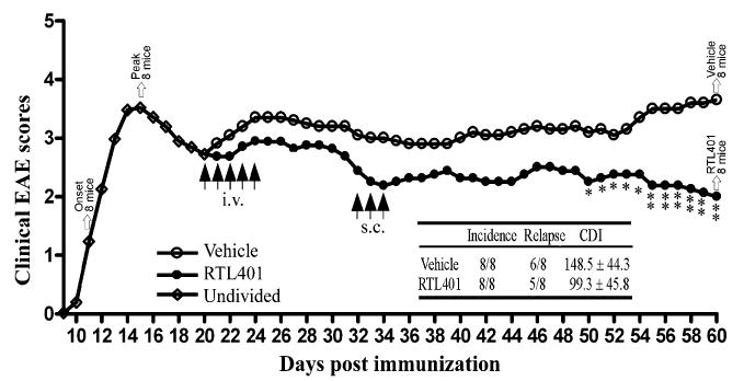
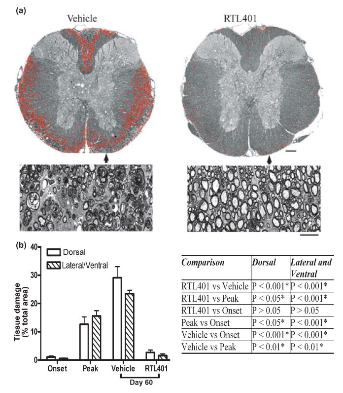
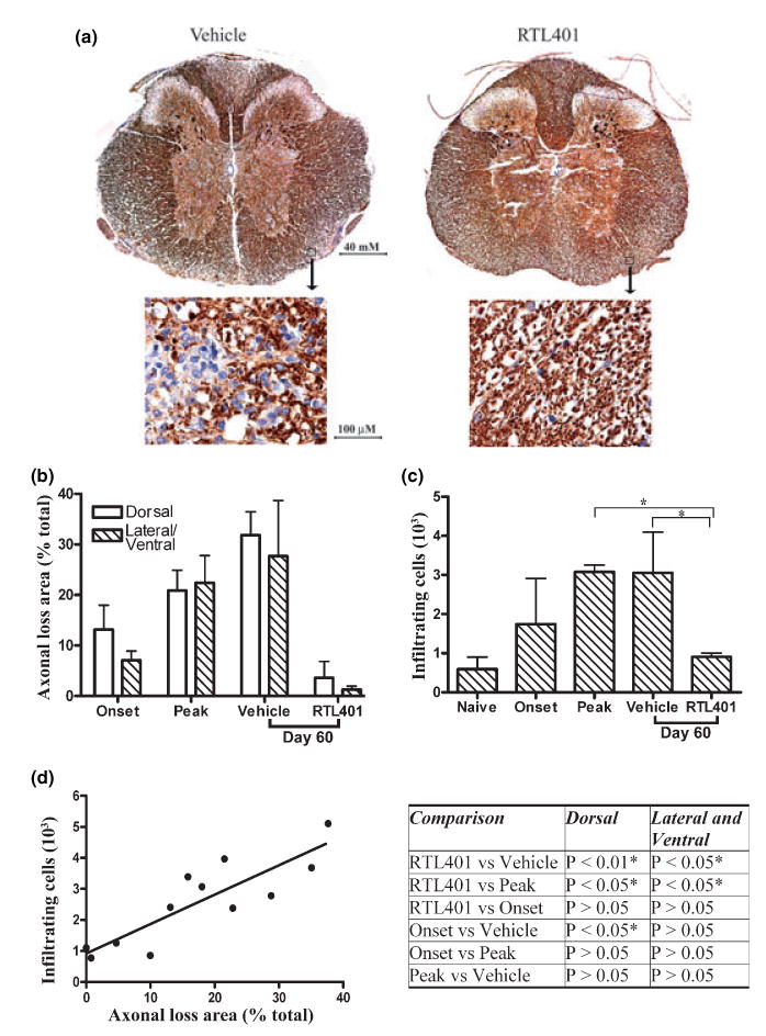
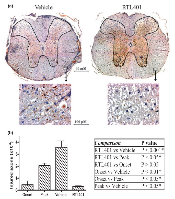
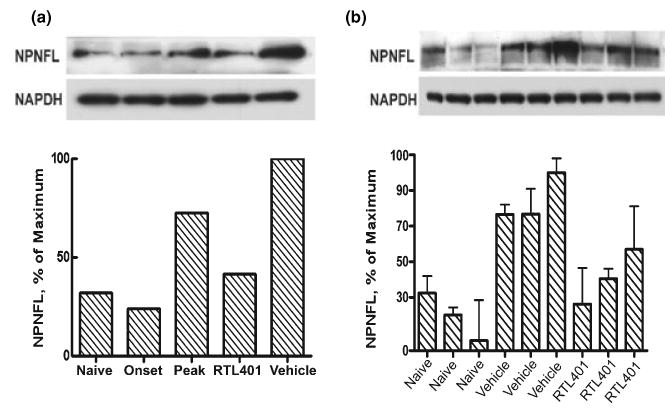
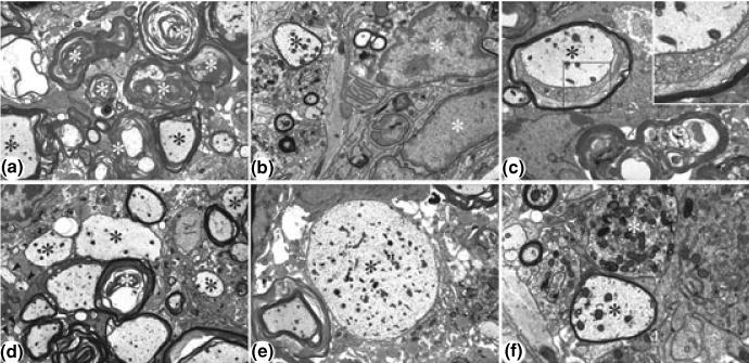
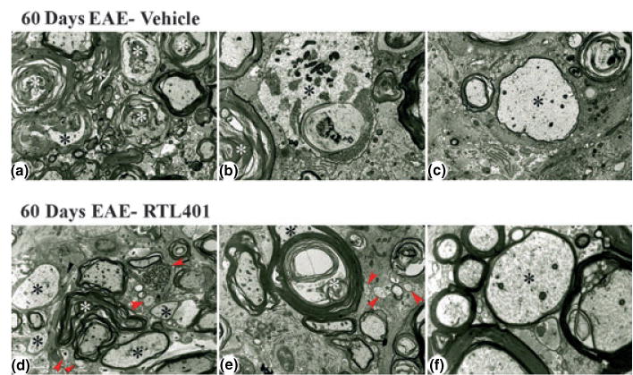
Inflammation results in CNS damage in multiple sclerosis (MS) and experimental autoimmune encephalomyelitis (EAE), an animal model of MS. It is uncertain how much repair of injured myelin and axons can occur following highly selective anti-inflammatory therapy in EAE and MS. In this study, SJL/J mice with established EAE were treated successfully with an antigen-specific recombinant T cell receptor ligand (RTL), RTL401, a mouse I-A(s)/PLP-139-151 construct, after the peak of EAE. To define the mechanisms by which late application of RTL401 inhibits EAE, we evaluated mice at different time points to assess the levels of neuroinflammation and myelin and axon damage in their spinal cords. Our results showed that RTL401 administered after the peak of acute EAE induced a marked reduction in inflammation in the CNS, associated with a significant reduction of demyelination, axonal loss and ongoing damage. Electron microscopy showed that RTL-treated mice had reduced pathology compared with mice treated with vehicle and mice at the peak of disease, as demonstrated by a decrease in continued degeneration, increase in remyelinating axons and the presence of an increased number of small, presumably regenerative axonal sprouts. These findings indicate that RTL therapy targeting encephalitogenic T cells may promote CNS neuroregenerative processes.


Figures

Fig. 1
Fig. 1
RTL401 treatment of SJL mice with established clinical EAE. Active EAE was induced in 32 female SJL mice by immunization with PLP-139–151 peptide/CFA. Eight mice (Onset) were euthanized at disease onset and another 8 (Peak) were euthanized at the peak of disease. On day 20, just after the peak of the first episode of EAE, the remaining mice were divided into two groups with matched disease scores (2.8 ± 0.7 for vehicle-treated mice vs 2.6 ± 0.5 for RTL401 treated mice) and treated daily for 5 days i.v. with RTL401 (100 μg/mouse) or vehicle. On day 32, the two groups of mice were boosted with 3 daily s.c. injections of RTL401 or vehicle and monitored for change of disease scores until the conclusion of the experiment (day 60). Inserted table: The effect of RTL401 treatment on cumulative disease index (CDI) and number of relapses of SJL mice with EAE. Data represent averages of summed daily disease scores for all available mice. Statistically significant difference in daily or collective EAE scores between RTL401- vs. vehicle-treated mice is demonstrated by Student t-test, p < 0.05* or 0.01**.
Fig. 2
Fig. 2
RTL treatment ameliorated tissue (myelin) damage in EAE. (a) Representative thoracic spinal cord sections from vehicle (left panel) or RTL401 (right panel)-treated mice stained with toluidine blue for myelin on day 60 of EAE. (b) Morphometric analysis of the development of myelin damage in the white matter of thoracic spinal cords of EAE treated with vehicle or RTL401. The cords were dissected from paraformaldehyde (PFA) plus glutaraldehyde-perfused EAE mice (4 mice randomly chosen from a group of 8, the rest were euthanized by perfusion with PBS, see Figs 3,4 and 5) euthanized at disease onset (day 11, disease score 1.5), peak (between day 15 and day 20 when the disease score for each mouse reached 4.5), or at the termination of the experiment (day 60 of EAE, after RTL401 or vehicle treatment). Tissue sections were stained with toluidine blue and images were captured with a compound microscope equipped with a digital camera. Areas with tissue damage were measured and analyzed using BIOQUANT classic 95 software. Scale bars = 25 μM (low power views) or 100 μM (high power views). Inserted Table: RTL401-treated mice show a significant reduction in spinal cord white matter tissue damage, demonstrated by one-way ANOVA followed by Newman-Kuels multiple comparisons test. *Comparison statistically significant.
Fig. 3
Fig. 3
RTL401-treatment decreased axonal loss and inflammation in the spinal cord in EAE. (a) Representative phosphorylated neuro-filament staining of axons in thoracic spinal cord sections from EAE mice treated with vehicle (left panel) or RTL401 (right panel) 60 days after disease induction. Tissue sections were dissected from PBS-perfused mice (4 available mice from a group of eight after 4 randomly chosen mice were perfused by PFA, see Fig. 2), fixed and stained with SMI312, an antibody cocktail for neurofilaments (brown). The nuclei of inflammatory mononuclear cells were visualized by hematoxylin (blue). Images were captured with a compound microscope equipped with a digital camera. (b) Morphometric analysis of the development of axonal loss in EAE mice treated with vehicle or RTL401. EAE mice were euthanized at disease onset (day 11, disease score 1.5), peak (between day 15 and day 20, when disease scores reached 4.5), or at the termination of the experiment (day 60 of EAE, after RTL401 or vehicle treatment). Digitally acquired images were analyzed with BIOQUANT software. Areas with loss of phosphorylated NF staining of axons were circled by hand and traced by BIOQUANT. The percentage of axonal loss area was calculated by dividing total axonal loss areas by the total area of dorsal or lateral/ventral white matter. Data represent mean ± SD (n = 4). (c) RTL401-treatment reduced inflammatory mononuclear cells in the CNS of EAE mice. Total numbers of inflammatory mononuclear cells (stained blue with hematoxylin) in whole thoracic spinal cord sections were counted manually. Data represent mean ± SD (n = 4). *Comparison statistically significant as demonstrated by one-way ANOVA followed by Newman-Kuels multiple comparisons test, n = 4. (d) Correlation of axonal loss to number of inflammatory cells in individual mice. Pearson’s correlation analysis showed a significant correlation, r = 0.8636, p (two-tailed) = 0.0003. Inserted Table: RTL401-treated mice show a significant reduction in axonal loss, demonstrated by one-way ANOVA followed by Newman-Kuels multiple comparisons test. *Comparison statistically significant.
Fig. 4
Fig. 4
RTL401-treatment reduced axonal injury in the spinal cord of EAE mice. (a) Representative non-phosphorylated neurofilaments (NPNFL), which are abnormally expressed in injured axons, of the thoracic spinal cord from EAE mice treated with vehicle (left panel) or RTL401 (right panel) 60 days after disease induction. Tissue sections were dissected from 4 mice per group, fixed and stained with antibody SMI32 for NPNFL (brown). The nuclei of inflammatory mononuclear cells were stained with hematoxylin (blue). Images were captured with a compound microscope equipped with a digital camera. (b) Morphometric analysis of the total number of injured (NPNFL-positive) axons in the whole thoracic spinal cord white matter in EAE mice. EAE mice were euthanized by perfusion with PBS at disease onset (day 11, disease score 1.5), peak (between day 15 and day 20, when disease scores reached 4.5), or at the termination of the experiment (day 60 of EAE, after RTL401 or vehicle treatment). The numbers of injured axons were counted manually by an investigator with no knowledge of treatment conditions. Data = mean ± SD (n = 4). Inserted Table: RTL401-treated mice show a significant reduction in the number of injured axons, demonstrated by one-way ANOVA followed by Newman-Kuels multiple comparisons test. *Comparison statistically significant.
Fig. 5
Fig. 5
RTL treatment ongoing axonal injury in the lumbar spinal cord of EAE mice. Left panel: Immunoblotting result showing that RTL401 treatment reversed the development of axonal injury as indicated by abnormal expression of non-phosphorylated neurofilaments in lumbar spinal cords from mice with EAE. EAE mice were euthanized by perfusion with PBS at disease onset (day 11, disease score 1.5), peak (between day 15 and day 20, when disease scores reached 4.5), or at the termination of the experiment (day 60 of EAE, after RTL401 or vehicle treatment). The lysates of whole lumbar spinal cords from each group (4 PBS-perfused mice from a group of 8) were pooled and the amount of NPNFL was detected after immunoblotting. Each column represents pooled samples from 4 mice from a single group. Right panel: Immunoblotting results showed the amount of NPNFL in the lumbar spinal cord samples from three randomly chosen mice (3 out of the 4 samples in the pool) from naïve, vehicle-treated (day 60 of EAE) or RTL401-treated (day 60 of EAE) groups. Each band represents a lumbar spinal cord sample from an individual mouse. The experiment was repeated two times.
Fig. 6
Fig. 6
Representative electron micrographs showing lesion areas in spinal cords from EAE mice at the peak of the disease. (a) Low power view of typical lesion area. Wallerian-like axonal degeneration (white asterisks) is the most prominent feature. Active demyelination (black asterisks) is also present. Magnification × 4000. (b) Inflammatory cells (white asterisks) are also present, enlarging the lesion area. Note remyelinating axon (black asterisk). Magnification × 8000. (c) Active demyelination (black asterisk), as revealed by the loosening of the myelin sheath. Inset: Higher power view of boxed region showing active demyelination. Magnification × 6700; × 14 000 (inset). (d) Low power view showing active demyelination (white asterisk), medium to large sized remyelinating axons (black asterisk) and several very small axons (arrowheads), presumably representing regenerating axonal sprouts. Magnification × 5000. (e) Low power view of a large, demyelinated axon (black asterisk). Magnification × 5000. (f) Higher power view of large remyelinating axon (black asterisk) and an end bulb of a degenerating axon (dystrophic axon) (white asterisk). Magnification × 14 000.
Fig. 7
Fig. 7
Representative electron micrographs showing lesion areas in spinal cords from mice with EAE on day 60 after treatment with vehicle (panels a–c) or RTL401 (panels d–f). (a) Low power view of typical lesion area showing marked continued Wallerian-like axonal degeneration (white asterisks) and demyelination (black asterisk). Note paucity of infiltrating cells and lack of small, regenerating axonal sprouts. Magnification × 4000. (b) Higher power view showing Wallerian-like axonal degeneration (white asterisk) and active demyelination (black asterisk). Magnification × 8000. (c) Higher power view of a large, remyelinating axon as shown by the relatively thin myelinated sheath (black asterisk). Magnification × 6700. (d) Low power view of typical lesion area showing continued Wallerian-like axonal degeneration (white asterisk), including a dystrophic axon (black arrow), and demyelinated and remyelinating axons (black asterisks). However, there are also prominent remyelinating axons and several small axonal sprouts (red arrowheads). Note paucity of infiltrating cells. Magnification × 4000. (e) Low power view of a large fiber (black asterisk) undergoing active demyelination (white asterisk). Note also three very small axons/regenerating sprouts (arrowheads). Magnification × 5000. (f) Higher power view of a medium-sized, remyelinating axon as shown by the relatively thin myelinated sheath (black asterisk). Magnification × 14 000.

Similar articles

Cited by

References

    1. Arnold DL, Matthews PM, Francis GS, O’Connor J, Antel JP. Proton magnetic resonance spectroscopic imaging for metabolic characterization of demyelinating plaques. Ann Neurol. 1992;31:235–241. - PubMed
    1. Back SA, Tuohy TM, Chen H, Wallingford N, Craig A, Struve J, Luo NL, Banine F, Liu Y, Chang A, Trapp BD, Bebo BF, Jr, Rao MS, Sherman LS. Hyaluronan accumulates in demyelinated lesions and inhibits oligodendrocyte progenitor maturation. Nat Med. 2005;11:966–972. - PubMed
    1. Barkhof F, Bruck W, De Groot CJ, Bergers E, Hulshof S, Geurts J, van Polman CH, van Polman dV. Remyelinated lesions in multiple sclerosis: magnetic resonance image appearance. Arch Neurol. 2003;60:1073–1081. - PubMed
    1. Bruck W. The pathology of multiple sclerosis is the result of focal inflammatory demyelination with axonal damage. J Neurol. 2005;252:v3–v9. - PubMed
    1. Burrows GG, Adlard KL, Bebo BF, Jr, Chang JW, Tenditnyy K, Vandenbark AA, Offner H. Regulation of encephalitogenic T cells with recombinant TCR ligands. J Immunol. 2000;164:6366–6371. - PubMed

Publication types

MeSH terms