. 2006 Oct 13;281(41):30725-35.
doi: 10.1074/jbc.M605814200. Epub 2006 Aug 9.

Mutant analysis of the Shal (Kv4) voltage-gated fast transient K+ channel in Caenorhabditis elegans

Affiliations

Mutant analysis of the Shal (Kv4) voltage-gated fast transient K+ channel in Caenorhabditis elegans

Gloria L Fawcett et al. J Biol Chem. .

Abstract

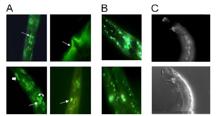
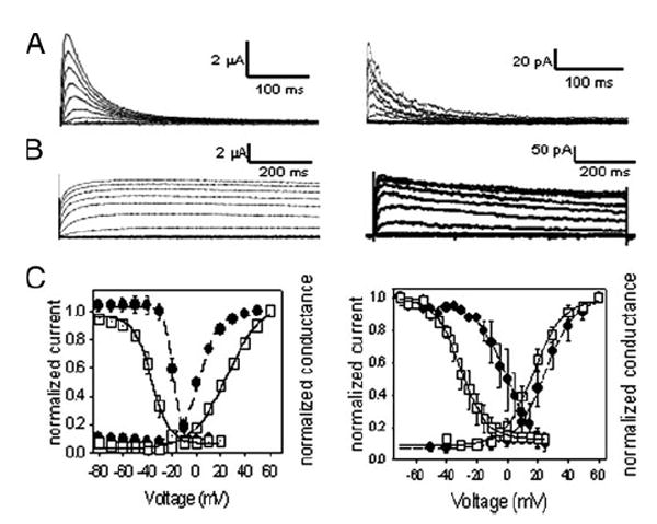
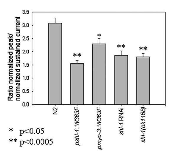
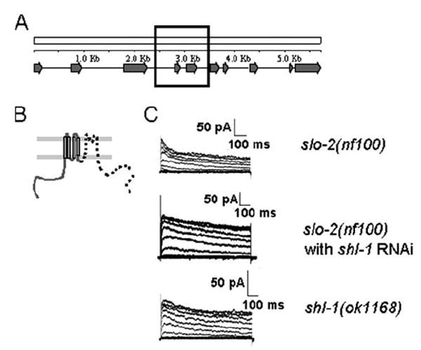
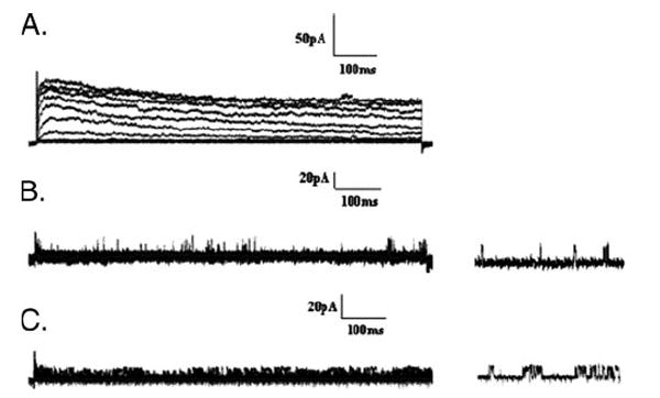
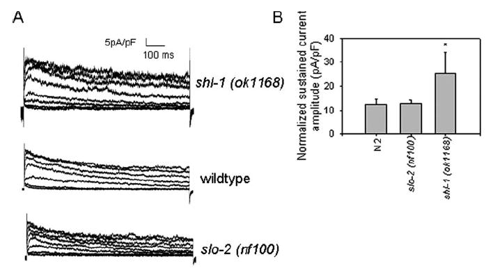
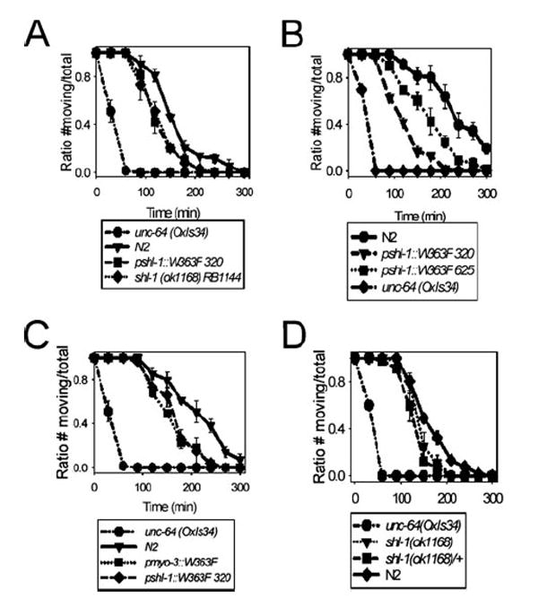
Shal (Kv4) alpha-subunits are the most conserved among the family of voltage-gated potassium channels. Previous work has shown that the Shal potassium channel subfamily underlies the predominant fast transient outward current in Drosophila neurons (Tsunoda, S., and Salkoff, L. (1995) J. Neurosci. 15, 1741-1754) and the fast transient outward current in mouse heart muscle (Guo, W., Jung, W. E., Marionneau, C., Aimond, F., Xu, H., Yamada, K. A., Schwarz, T. L., Demolombe, S., and Nerbonne, J. M. (2005) Circ. Res. 97, 1342-1350). We show that Shal channels also play a role as the predominant transient outward current in Caenorhabditis elegans muscle. Green fluorescent protein promoter experiments also revealed SHL-1 expression in a subset of neurons as well as in C. elegans body wall muscle and in male-specific diagonal muscles. The shl-1 (ok1168) null mutant removed all fast transient outward current from muscle cells. SHL-1 currents strongly resembled Shal currents in other species except that they were active in a more depolarized voltage range. We also determined that the remaining delayed-rectifier current in cultured myocytes was carried by the Shaker ortholog SHK-1. In shl-1 (ok1168) mutants there was a significant compensatory increase in the SHK-1 current. Male shl-1 (ok1168) animals exhibited reduced mating efficiency resulting from an apparent difficulty in locating the hermaphrodite vulva. SHL-1 channels are apparently important in fine-tuning complex behaviors, such as mating, that play a crucial role in the survival and propagation of the species.


Figures

FIGURE 1
FIGURE 1. Cell-type expression patterns of shl-1 and shk-1
A, GFP expression of the transgene pshl-1∷shl-1∷GFP in N2 animals. Expression was observed in a variety of neurons, including pharyngeal neurons (top left, white arrow), interneurons (bottom left, thin arrow), sensory neurons (bottom left, curved arrow), and phasmid neurons (bottom right, white arrow). In addition, we observed expression in two muscle types, body wall muscle (bottom left, thick arrow) and vulval muscle (top right, white arrow). B, GFP expression of the transgene pshk-1∷shk-1∷GFP in N2 animals. Expression was observed in a variety of neurons in the ganglia of the head (top). Expression throughout body wall muscle cells was visible (bottom) in a striated pattern. C, top, GFP expression of the transgene pshl-1∷shl-1∷GFP in male N2 animals. Expression was observed in the eight male-specific diagonal muscle cells. C, bottom, Nomarski picture of GFP view above.
FIGURE 2
FIGURE 2. Comparison of conductance voltage and steady-state inactivation properties of SHK-1 and SHL-1 in C. elegans slo-2 (nf100) myocytes or heterologously expressed in Xenopus oocytes
A, left, currents of SHL-1 heterologously expressed in Xenopus oocytes were obtained under voltage clamp at a holding potential of −100 mV with 10-mV steps from −80 to +50 mV for 450 ms with 5-s interpulse intervals. A, right, whole-cell currents from shk-1 RNAi treated slo-2(nf100) myocytes were obtained under voltage clamp at a holding potential of −70 mV with 10-mV steps from −70 to +60 mV with 5-s interpulse intervals. B, left, currents from SHK-1 isoform a heterologously expressed in Xenopus oocytes obtained under voltage clamp at a holding potential of −100 mV with 10-mV steps from −80 to +60 mV. B, right, currents from shl-1(ok1168) myocytes were obtained under voltage clamp at a holding potential of −70 mV with 10-mV steps from −70 mV to +60 mV with 5-s interpulse intervals. C, left, steady-state inactivation and activation data plotted for SHL-1 and SHK-1 heterologously expressed in Xenopus oocytes. □, SHL-1; ■, SHK-1. Best fits for Boltzmann equation are shown as lines through data points. V0.5a = 25.0±1.6 mV (n = 7), ka = 14. 0±1.2 mV;V0.5i = −36.7 ± 0.7 mV (n = 7), ki = 7. 3 ± 0.6 mV. V0.5a = 2. 4 ± 0.8 mV (n = 8), ka = 7. 4 ± 0.7 mV; V0.5i = −19. 5 ± 2.8 mV (n = 8), ki = 3. 7 ± 0.3 mV. Calculated conductance used the equation G = I/(EEK), where G is the conductance; I is the measured current; E is the test voltage, and EK is estimated at −96 mV. Steady-state inactivation data were obtained using a voltage step protocol stepping from −120 to +20 mV in 10-mV steps, with a holding potential of −100 mV. Normalization was obtained by the following ratio: I/Imax, where Imax is the maximum observed current, and I is the current at any given voltage. C, right, steady-state inactivation and activation data were plotted for SHL-1 and SHK-1 currents observed in cultured C. elegans myocytes. The activation parameter values were V0. 5a = 11.2 ± 1.5 mV, ka = 14.1 ± 1.04 (n = 5); V0.5a = 20. 4 ± 2, ka = 7. 7 ± 1.1 for SHL-1 (□)(n = 3) and SHK-1 (■) (n = 7) currents, respectively. SHL-1 currents inactivated with values of V0.5i = −33.1 mV ± 1.2 and ki = 8.3 ± 0.7 (n = 2), whereas SHK-1 inactivation parameter values were V0. 5i = −6.95±1.7, ki = 5.8±0.5 (n =3). Internal concentrations of Cl and Ca2+ were 120 mm and 10 nm, respectively. Previously published in Santi et al. (7).
FIGURE 3
FIGURE 3. Removal of SHL-1 current in cultured C. elegans myocytes
The ratio of the peak current (pA/pF) to the minimum sustained current (pA/pF) is plotted for the wild type N2 strain (3.0831 ± 0.1973) (n = 7), the dominant negative strains pshl-1∷W363F (1. 5623±0. 1101) (n=5) and pmyo-3∷W363F (2.2910±0.0020) (n=5), shl-1RNAi-treated myocytes (1.5723±0.1502) (n=3), and the deletion mutant shl-1(ok1168) (1.8088±0.1324) (n=6). * signifies p < 0.05. ** signifies p < 0.0005.
FIGURE 4
FIGURE 4. Characterization of the shl-1 (ok1168) deletion mutant breakpoints
A, schematic of shl-1 genomic locus in C. elegans showing all 10 exons of the gene. The black box represents the genomic area that is deleted in the shl-1(ok1168) deletion mutant. B, pictorial representation of the truncated protein that might be produced by the shl-1(ok1168) mutant. The deletion removes the terminal 3 amino acids of the fourth transmembrane domain, and a frameshift resulting from the splicing of exons 3 and 6 results in missense coding of 16 amino acids ending in a stop codon. The portion of the channel that has been removed is designated by a dashed line. C, slo-2(nf100) myocytes display two voltage-sensitive macroscopic outward currents because of the presence of SHL-1 and SHK-1 channels (n = 7) (top). RNAi removal of SHL-1 current results in retention of a slowly inactivating voltage-sensitive current, SHK-1 (n = 3) (middle). The shl-1(ok1168) deletion mutant (n= 7) results in removal of fast transient current (bottom). Cells were held at −70 mV and stepped from −70 to +60 mV in 10-mV increments.
FIGURE 5
FIGURE 5. Removal of all macroscopic outward currents in C. elegans myocytes
A, currents from slo-2(nf100) cells are shown as a control where both SHL-1 and SHK-1 currents are present. Whole-cell recordings were obtained from a holding potential of −70 mV, and stepped from −70 to +60 mV in 10-mV intervals. B, dominant negative pshl-1∷shl-1∷W363F cells (n = 3) treated with shk-1 RNAi to remove both SHL-1 and SHK-1 macroscopic currents. SLO-2 currents were inhibited by limiting the intracellular Ca2+ and Cl. There is a complete removal of all macroscopic SHL-1 and SHK-1 currents in these myocytes. Unidentified single channel openings can be seen in these records (right traces). C, shl-1(ok1168) deletion mutant cells (n = 3) treated with shk-1 RNAi as in B.
FIGURE 6
FIGURE 6. Up-regulation of SHK-1 current in shl-1 (ok1168) mutant myocytes
A, current traces from shl-1 (ok1168) myocytes (top), N2 wild-type myocytes (middle), and slo-2 (nf100) myocytes (bottom) shown on the same scale normalized for cell capacitance. B, mean normalized sustained current amplitudes with standard errors for the slo-2 (nf100) myocytes (12.5 pA/pF±1.9), N2 myocytes (12.6 pA/pF±1.5), and the shl-1 (ok1168) myocytes (25.5 pA/pF ± 8.5). * indicates p < 0.05 compared with shl-1 (ok1168) myocytes.
FIGURE 7
FIGURE 7. Removal of SHL-1 current in body wall muscle is sufficient to confer aldicarb hypersensitivity with 0.25 mm aldicarb
A, both the shl-1(ok1168) deletion mutant and the pshl-1∷W363F∷GFP line 320 carrying the dominant negative construct displayed aldicarb hypersensitivity compared with N2 animals. B, we tested two lines carrying dominant negative constructs integrated on different chromosomes to ensure that the observed aldicarb hypersensitivity phenotype was not because of position effects in these transgenic animals. Both lines carrying integrated constructs (320 on chromosome X and 625 on chromosome IV) displayed aldicarb hypersensitivity. The aldicarb hypersensitivity of the dominant negative transgenic line was not influenced by position effects. C, expression of the dominant negative transgene under control of pmyo-3 (a muscle-specific promoter) resulted in aldicarb hypersensitivity similar to that observed for the dominant negative under the native shl-1 promoter. D, the heterozygous shl-1(ok1168)/+ animals had an intermediate phenotype which fell between N2 and shl-1(ok1168) homozygotes. The positive control for hypersensitivity to aldicarb for all assays was the strain unc-64(OxIs34), a gain-of-function integrated transgenic syntaxin line.

Similar articles

Cited by

References

    1. Tsunoda S, Salkoff L. J Neurosci. 1995;15:1741–1754. - PMC - PubMed
    1. Guo W, Jung WE, Marionneau C, Aimond F, Xu H, Yamada KA, Schwarz TL, Demolombe S, Nerbonne JM. Circ Res. 2005;97:1342–1350. - PubMed
    1. Hagiwara S, Kusano K, Saito N. J Physiol (Lond) 1961;155:471–489. - PMC - PubMed
    1. Connor JA, Stevens CF. J Physiol (Lond) 1971;213:21–30. - PMC - PubMed
    1. Neher E. J Gen Physiol. 1971;58:36–53. - PMC - PubMed

Publication types

LinkOut - more resources