

Up-regulation of transient receptor potential canonical 1 (TRPC1) following sarco(endo)plasmic reticulum Ca2+ ATPase 2 gene silencing promotes cell survival: a potential role for TRPC1 in Darier's disease
- PMID: 16899508
- PMCID: PMC1635355
- DOI: 10.1091/mbc.e06-03-0251
Up-regulation of transient receptor potential canonical 1 (TRPC1) following sarco(endo)plasmic reticulum Ca2+ ATPase 2 gene silencing promotes cell survival: a potential role for TRPC1 in Darier's disease
Abstract
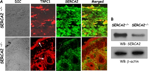
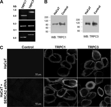
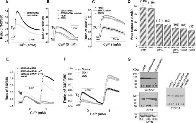
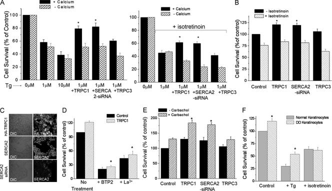
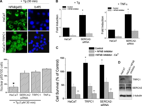
The mechanism(s) involved in regulation of store operated calcium entry in Darier's disease (DD) is not known. We investigated the distribution and function of transient receptor potential canonical (TRPC) in epidermal skin cells. DD patients demonstrated up-regulation of TRPC1, but not TRPC3, in the squamous layers. Ca2+ influx was significantly higher in keratinocytes obtained from DD patients and showed enhanced proliferation compared with normal keratinocytes. Similar up-regulation of TRPC1 was also detected in epidermal layers of SERCA2+/- mice. HaCaT cells expressed TRPC1 in the plasma membrane. Expression of sarco(endo)plasmic reticulum Ca2+ ATPase (SERCA)2 small interfering RNA (siRNA) in HaCaT cells increased TRPC1 levels and thapsigargin-stimulated Ca2+ influx, which was blocked by store-operated calcium entry inhibitors. Thapsigargin-stimulated intracellular Ca2+ release was decreased in DD cells. DD keratinocytes exhibited increased cell survival upon thapsigargin treatment. Alternatively, overexpression of TRPC1 or SERCA2-siRNA in HaCaT cells demonstrated resistance to thapsigargin-induced apoptosis. These effects were dependent on external Ca2+ and activation of nuclear factor-kappaB. Isotretinoin reduced Ca2+ entry in HaCaT cells and decreased survival of HaCaT and DD keratinocytes. These findings put forward a novel consequence of compromised SERCA2 function in DD wherein up-regulation of TRPC1 augments cell proliferation and restrict apoptosis. We suggest that the anti-apoptotic effect of TRPC1 could potentially contribute to abnormal keratosis in DD.
Figures


Similar articles
-
ER-to-Golgi blockade of nascent desmosomal cadherins in SERCA2-inhibited keratinocytes: Implications for Darier's disease.Traffic. 2017 Apr;18(4):232-241. doi: 10.1111/tra.12470. Epub 2017 Feb 28. Traffic. 2017. PMID: 28156030 Free PMC article.
-
Darier's disease: a calcium-signaling perspective.Cell Mol Life Sci. 2008 Jan;65(2):205-11. doi: 10.1007/s00018-007-7397-z. Cell Mol Life Sci. 2008. PMID: 18049860 Free PMC article. Review.
-
Impaired trafficking of the desmoplakins in cultured Darier's disease keratinocytes.J Invest Dermatol. 2003 Dec;121(6):1349-55. doi: 10.1046/j.1523-1747.2003.12557.x. J Invest Dermatol. 2003. PMID: 14675181
-
Expression of sarco/endo-plasmic reticulum Ca2+-ATPase type 2 isoforms (SERCA2) in normal human skin and mucosa, and Darier's disease skin.Br J Dermatol. 2002 Oct;147(4):670-4. doi: 10.1046/j.1365-2133.2002.04916.x. Br J Dermatol. 2002. PMID: 12366411
-
Markers of squamous cell carcinoma in sarco/endoplasmic reticulum Ca2+ ATPase 2 heterozygote mice keratinocytes.Prog Biophys Mol Biol. 2010 Sep;103(1):81-7. doi: 10.1016/j.pbiomolbio.2009.10.005. Epub 2009 Oct 17. Prog Biophys Mol Biol. 2010. PMID: 19840814 Review.
Cited by
-
Transient receptor potential canonical 3 (TRPC3) mediates thrombin-induced astrocyte activation and upregulates its own expression in cortical astrocytes.J Neurosci. 2010 Sep 29;30(39):13116-29. doi: 10.1523/JNEUROSCI.1890-10.2010. J Neurosci. 2010. PMID: 20881130 Free PMC article.
-
TRPC1 contributes to light-touch sensation and mechanical responses in low-threshold cutaneous sensory neurons.J Neurophysiol. 2012 Feb;107(3):913-22. doi: 10.1152/jn.00658.2011. Epub 2011 Nov 9. J Neurophysiol. 2012. PMID: 22072513 Free PMC article.
-
The TRPC1 Ca2+-permeable channel inhibits exercise-induced protection against high-fat diet-induced obesity and type II diabetes.J Biol Chem. 2017 Dec 15;292(50):20799-20807. doi: 10.1074/jbc.M117.809954. Epub 2017 Oct 26. J Biol Chem. 2017. PMID: 29074621 Free PMC article.
-
ER-to-Golgi blockade of nascent desmosomal cadherins in SERCA2-inhibited keratinocytes: Implications for Darier's disease.Traffic. 2017 Apr;18(4):232-241. doi: 10.1111/tra.12470. Epub 2017 Feb 28. Traffic. 2017. PMID: 28156030 Free PMC article.
-
Patients with Darier disease have an increased risk of keratinocyte carcinoma: a Swedish registry-based nationwide cohort study.Orphanet J Rare Dis. 2024 Dec 16;19(1):463. doi: 10.1186/s13023-024-03497-z. Orphanet J Rare Dis. 2024. PMID: 39681873 Free PMC article.
References
-
- Ahn W., Lee M. G., Kim K. H., Muallem S. Multiple effects of SERCA2b mutations associated with Darier's disease. J. Biol. Chem. 2003;278:20795–20801. - PubMed
-
- Anglade P., Vyas S., Javoy-Agid F., Herrero M. T., Michel P. P., Marquez J., Mouatt-Prigent A., Ruberg M., Hirsch E. C., Agid Y. Apoptosis and autophagy in nigral neurons of patients with Parkinson's disease. Histol. Histopathol. 1997;12:25–31. - PubMed
-
- Berridge M. J., Lipp P., Bootman M. D. The versatility and universality of calcium signalling. Nat. Rev. Mol. Cell Biol. 2000;1:11–21. - PubMed
-
- Benzaquen L. R., Brugnara C., Byers H. R., Cattoni-Celli S. Clotrimazole inhibits cell proliferation in vitro and in vivo. Nat. Med. 1995;6:534–537. - PubMed
Publication types
MeSH terms
Substances
Grants and funding
LinkOut - more resources
Full Text Sources
Other Literature Sources
Molecular Biology Databases
Miscellaneous
