. 2006 Oct;17(10):4420-34.
doi: 10.1091/mbc.e06-03-0245. Epub 2006 Aug 9.

Gamma-tubulin is required for proper recruitment and assembly of Kar9-Bim1 complexes in budding yeast

Affiliations

Gamma-tubulin is required for proper recruitment and assembly of Kar9-Bim1 complexes in budding yeast

Lara Cuschieri et al. Mol Biol Cell. 2006 Oct.

Abstract

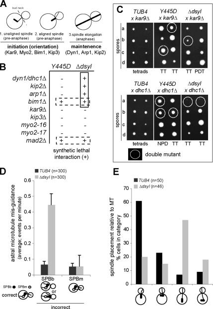
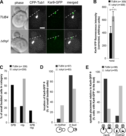
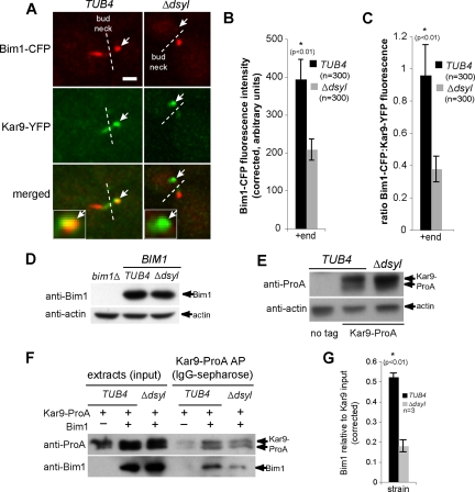
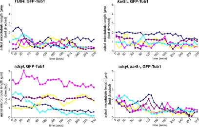
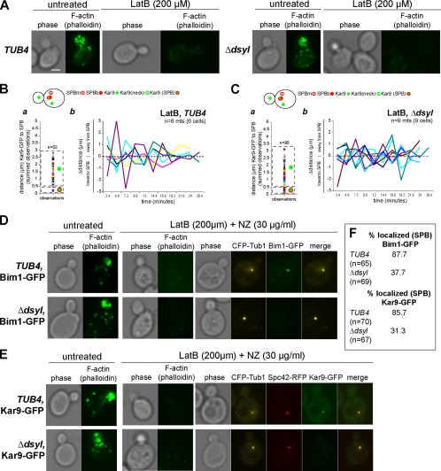
Microtubule plus-end-interacting proteins (+TIPs) promote the dynamic interactions between the plus ends (+ends) of astral microtubules and cortical actin that are required for preanaphase spindle positioning. Paradoxically, +TIPs such as the EB1 orthologue Bim1 and Kar9 also associate with spindle pole bodies (SPBs), the centrosome equivalent in budding yeast. Here, we show that deletion of four C-terminal residues of the budding yeast gamma-tubulin Tub4 (tub4-delta dsyl) perturbs Bim1 and Kar9 localization to SPBs and Kar9-dependent spindle positioning. Surprisingly, we find Kar9 localizes to microtubule +ends in tub4-delta dsyl cells, but these microtubules fail to position the spindle when targeted to the bud. Using cofluorescence and coaffinity purification, we show Kar9 complexes in tub4-delta dsyl cells contain reduced levels of Bim1. Astral microtubule dynamics is suppressed in tub4-delta dsyl cells, but it are restored by deletion of Kar9. Moreover, Myo2- and F-actin-dependent dwelling of Kar9 in the bud is observed in tub4-delta dsyl cells, suggesting defective Kar9 complexes tether microtubule +ends to the cortex. Overproduction of Bim1, but not Kar9, restores Kar9-dependent spindle positioning in the tub4-delta dsyl mutant, reduces cortical dwelling, and promotes Bim1-Kar9 interactions. We propose that SPBs, via the tail of Tub4, promote the assembly of functional +TIP complexes before their deployment to microtubule +ends.


Figures

Figure 1.
Figure 1.
Localization of Bim1 and Kar9 to SPBs is defective in tub4-Δdsyl cells. (A) TUB4 and tub4-Y445D cells treated with 30 μg/ml NZ had detectable Bim1–GFP foci associated with the collapsed spindle (single focus of CFP–Tub1), whereas the majority of tub4-Δdsyl cells treated with NZ did not. (B) Histogram showing the percentage of TUB4, tub4-Y445D, and tub4-Δdsyl cells with detectable Bim1–GFP foci at collapsed spindles after NZ treatment. (C) TUB4 and tub4-Y445D cells treated with NZ had Kar9–GFP foci localized to the SPBb. In contrast, the majority of tub4-Δdsyl cells lacked detectable Kar9–GFP foci associated with SPBs. Spc42–RFP marks the SPBb. Microtubules are labeled with CFP–Tub1. (D) Histogram indicating the percentage of TUB4, tub4-Y445D, and tub4-Δdsyl cells exhibiting Kar9–GFP foci at SPBs.
Figure 2.
Figure 2.
DSYL residues are required for function of the Kar9 pathway. (A) Cartoon of spindle positioning pathways in budding yeast. Early spindle placement is dependent on the Kar9 pathway, whereas the maintenance of spindle placement during anaphase requires the dynein pathway. (B) Query alleles (tub4-Y445D and tub4-Δdsyl) were analyzed for synthetic lethal interactions with mutations that disrupt either Kar9 or Dhc1 function. Synthetic lethal (+) and viable interactions (−) are indicated. The tub4-Δdsyl allele was synthetically lethal with mutations that disrupt Dhc1 function but not Kar9 function. (C) Representative tetrads for the analysis in B. Double mutant spores are circled. tub4-Δdsyl dhc1Δ double mutants failed to grow, whereas tub4-Δdsyl kar9Δ double mutants were viable. n = 40 tetrads for each cross. (D) The frequency of astral microtubule misguidance (visualized by GFP–Tub1) that occurred from the mother-bound SPB (SPBm) or SPBb was measured in TUB4 and tub4-Δdsyl cells. tub4-Δdsyl cells had a higher frequency of microtubule misguidance relative to wild-type. n = 300. (E) Analysis of spindle placement in TUB4 and tub4-Δdsyl cells. tub4-Δdsyl cells had a higher frequency of spindles that were positioned far from the bud neck relative to TUB4 cells. n = 300. Categories of spindle placement assessed are represented in the cartoon image.
Figure 3.
Figure 3.
Kar9 preferentially localizes to astral microtubule +ends in tub4-Δdsyl cells. (A) In tub4-Δdsyl cells, Kar9–GFP localized to the +ends of astral microtubules. Dashed line indicates the position of the bud neck. Bar, 2 μm. (B) Histogram indicating the fluorescent intensities of Kar9–GFP foci associated with microtubule +ends in TUB4 and tub4-Δdsyl cells. tub4-Δdsyl cells (n = 300; p < 0.01) have a higher average fluorescent intensity of Kar9–GFP associated with microtubule +ends relative to those of TUB4 (n = 300; p < 0.01) cells. (C) Quantification of Kar9 localization in TUB4 and tub4-Δdsyl cells. The majority of tub4-Δdsyl cells have Kar9–GFP foci associated with microtubule +ends. (D) Histogram indicating the localization of Kar9–GFP foci in TUB4 and tub4-Δdsyl cells. The majority of Kar9–GFP foci are located within the bud for both cell types. (E) Spindle positioning relative to Kar9–GFP foci was analyzed in TUB4 and tub4-Δdsyl cells. In tub4-Δdsyl cells, spindles remain mispositioned even when a Kar9-loaded astral microtubule is properly targeted into the bud. Categories of spindle placement assessed are represented in the cartoon image.
Figure 4.
Figure 4.
Stoichiometry of Bim1–Kar9 complexes is reduced in tub4-Δdsyl cells. (A) In TUB4 cells, Bim1–CFP and Kar9–YFP are observed on microtubule +ends (white arrows). In contrast, Kar9–YFP foci are detected on +ends lacking detectable Bim1–CFP (white arrows) in the majority of tub4-Δdsyl cells. White dashed line indicates bud neck. Bar, 2 μm. (B) Quantification of Bim1–CFP fluorescence indicated that the amount of Bim1 localized to microtubule +ends is significantly reduced in tub4-Δdsyl cells relative to wild-type (p < 0.01). (C) The ratio of Bim1–CFP/Kar9–YFP fluorescence is greater in TUB4 cells (0.958 ± 0.224 arbitrary fluorescent units) relative to tub4-Δdsyl cells (0.377 ± 0.078 arbitrary fluorescent units). (D and E) Bim1 and Kar9 protein levels in tub4-Δdsyl cells are similar to those in TUB4 cells. Actin acts as a loading control. (F) Bim1 copurifies with Kar9–ProA in wild-type and tub4-Δdsyl cells, but the level of Bim1 copurification is reduced in tub4-Δdsyl cells. (G) Histogram showing the averaged amount of copurifying Bim1 (arbitrary units normalized to the Kar9–ProA input) in three independent experiments for wild-type and tub4-Δdsyl strains.
Figure 5.
Figure 5.
Preferential localization of Kar9 on microtubule +ends in tub4-Δdsyl cells alters astral microtubule dynamics. Microtubule lifetime lengths were measured in TUB4, tub4-Δdsyl, kar9Δ, and tub4-Δdsyl kar9Δ cells containing a GFP–Tub1 fusion protein (see Materials and Methods for experimental details). Microtubule lengths (micrometers) were measured in triplicate at 10-s intervals (total time 310 s), and the averaged length was plotted relative to time (seconds). Astral microtubules in tub4-Δdsyl cells were less dynamic relative to TUB4 and kar9Δ cells with an increase in the duration of pause event. Dynamics are restored in tub4-Δdsyl kar9Δ double mutant cells.
Figure 6.
Figure 6.
Kar9 interactions with Myo2 are stabilized in tub4-Δdsyl cells. (A) In TUB4 cells microtubules loaded with Kar9 (green arrows) are dynamic and retract quickly from the cortex. In tub4-Δdsyl cells, Kar9-associated microtubules are less dynamic and rarely depolymerize back to the SPBb (red arrows) or bud neck (indicated by dashed line). Mother and bud are outlined (white) in the first frame of the montage. (B) In wild-type cells, Kar9–GFP foci distribute near and far from the SPBb (B, a). Kar9–GFP foci are highly dynamic and are associated with both growing and shrinking microtubules (B, b). In tub4-Δdsyl cells (C, a), the Kar9–GFP foci to not cluster near the SPBb but rather to a region ∼1.5–3 μm in length from the SPBb. Furthermore, Kar9–GFP foci are not dynamic and are associated with microtubules that do not move toward and away from the SPBb (C, b). (D) Montage (t < 30 min) of Kar9 foci in myo2-16 cells and tub4-Δdsyl myo2-16 double mutants revealed that Kar9 is dynamic in both cell types. Dashed line indicates bud neck. Bar, 2 μm. (E) In myo2-16 Kar9–GFP foci have a large distribution due to the growth and shrinkage of dynamic microtubules (E, a). Microtubules in the myo2-16 cells are dynamic due to short interactions with the cortex (E, b). (F) In myo2-16, tub4-Δdsyl double mutants, Kar9–GFP foci are distributed near the SPBb and the bud cortex (F, a), whereas Kar9 dynamics are restored (F, b).
Figure 7.
Figure 7.
Stable localization of Kar9 in the bud depends on cortical actin. (A) Actin staining of TUB4 and tub4-Δdsyl cells with and without treatment of 200 μM LatB. Treatment with LatB resulted in the depolymerization of the actin cytoskeleton in TUB4 and tub4-Δdsyl cells. Respective phase images are shown. Bar, 2 μm. (B, a) In wild-type cells treated with LatB, Kar9–GFP foci distributed to at the bud cortex as well as the SPB. After latrunculin treatment, microtubules (Kar9–GFP foci) in wild-type cells were highly dynamic. In tub4-Δdsyl cells treated with latrunculin, the distribution of Kar9–GFP foci relative to the SPB were rescued to near wild-type levels (C, a). Kar9 dynamics were also rescued to near wild-type levels (C, b). (D–F) Analysis of Bim1 and Kar9 SPB localization in cells treated with 200 μM LatB (to disrupt F-actin) and 30 μg/ml NZ (to depolymerize astral microtubules). CFP–Tub1 was used to visualize the collapsed spindle and Alexa 488-phallodin to confirm the disruption of the actin cytoskeleton in the presence or absence of latrunculin and NZ. The majority of wild-type cells (87.7%; n = 65) treated with LatB and NZ had detectable Bim1–GFP associated with the collapsed spindle (D and F). In contrast, the percentage of LatB and NZ treated tub4-Δdsyl cells with detectable Bim1–GFP at SPBs was reduced (37.7%; n = 69) (D and F). Similarly, the localization of Kar9–GFP in tub4-Δdsyl cells treated with LatB and NZ was reduced; 31.3% of tub4-Δdsyl cells (n = 67) had detectable Kar9–GFP compared with 78% of wild-type cells (n = 70; E and F).
Figure 8.
Figure 8.
Kar9 function is restored by overproduction of Bim1. (A) Percentage of viable tub4-Δdsyl Δdhc1 double mutants recovered in the presence of overproduced Bim1, Kar9 or vector control. Overproduction of Bim1 (pBIM1) rescued the synthetic lethality observed in tub4-ΔdsylΔdhc1 double mutants, whereas overproduction of Kar9 (pKAR9) or expression of the vector control (pRS423) did not rescue synthetic lethality. Forty tetrads/mating were scored. (B) Bim1 and Kar9 interactions are restored by overproduction of Bim1. The level of Bim1 that coaffinity purifies with Kar9 in tub4-Δdsyl cells is restored in the presence of overproduced Bim1 relative to cells containing the vector control. (C) Astral microtubule guidance is restored by an overproduction of Bim1. tub4-Δdsyl pBIM1 cells have a lower occurrence of microtubule misguidance relative to tub4-Δdsyl pRS423 cells. (D) Spindle placement was analyzed in tub4-Δdsyl pRS423 and tub4-Δdsyl pBIM1 cells. Overproduction of Bim1 (black bars) rescued spindle positioning defects observed in control cells (gray bars).
Figure 9.
Figure 9.
Overproduction of Bim1 restores Kar9 dynamics in tub4-Δdsyl cells. (A) Montage of Kar9–GFP movements in tub4-Δdsyl pRS423 and tub4-Δdsyl pBIM1 cells. Overproduction of Bim1 rescues the Kar9 associated microtubules dynamics in tub4-Δdsyl cells, whereas in the vector control, Kar9-associated microtubules remain un-dynamic (green arrows). (B and C) Microtubules/Kar9–GFP foci were tracked during a time course (t < 30 min) in tub4-Δdsyl pRS423 and tub4-Δdsyl pBIM1 cells. Line graphs depict the length from the microtubule +ends relative to the SPBb per Δtime (t = 2.4 min). Scatter plots display the relative distribution of Kar9–GFP foci relative to the SPB over time. In tub4-Δdsyl pRS423 cells (B, a), the Kar9–GFP foci cluster to a region ∼1.5–3 μm in length from the SPBb, indicating the microtubules do not depolymerize back to the SPBb. Microtubules seemed to be static (B, b). In contrast, in tub4-Δdsyl cells with overproduced Bim1, the distribution of Kar9–GFP foci relative to the SPBb (C, a) and its dynamic movement in the bud (C, b) were similar to wild type.

Similar articles

Cited by

References

    1. Adames N. R., Cooper J. A. Microtubule interactions with the cell cortex causing nuclear movements in Saccharomyces cerevisiae. J. Cell Biol. 2000;149:863–874. - PMC - PubMed
    1. Aldaz H., Rice L. M., Stearns T., Agard D. A. Insights into microtubule nucleation from the crystal structure of human γ-tubulin. Nature. 2005;435:523–527. - PubMed
    1. Beach D. L., Thibodeaux J., Maddox P., Yeh E., Bloom K. The role of the proteins Kar9 and Myo2 in orienting the mitotic spindle of budding yeast. Curr. Biol. 2000;10:1497–1506. - PubMed
    1. Carminati J. L., Stearns T. Microtubules orient the mitotic spindle in yeast through dynein-dependent interactions with the cell cortex. J. Cell Biol. 1997;138:629–641. - PMC - PubMed
    1. Christianson T. W., Sikorski R. S., Dante M., Shero J. H., Hieter P. Multifunctional yeast high-copy-number shuttle vectors. Gene. 1992;110:119–122. - PubMed

Publication types

MeSH terms