

LC-MS/MS analysis of apical and basolateral plasma membranes of rat renal collecting duct cells
- PMID: 16899541
- PMCID: PMC2412072
- DOI: 10.1074/mcp.M600177-MCP200
LC-MS/MS analysis of apical and basolateral plasma membranes of rat renal collecting duct cells
Abstract
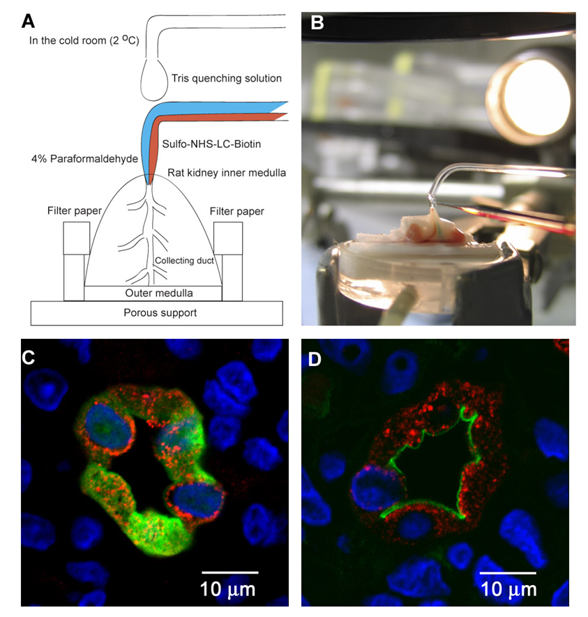
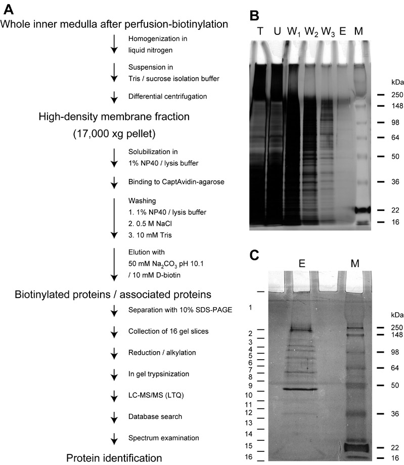
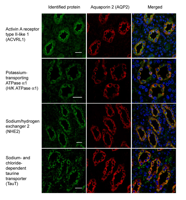
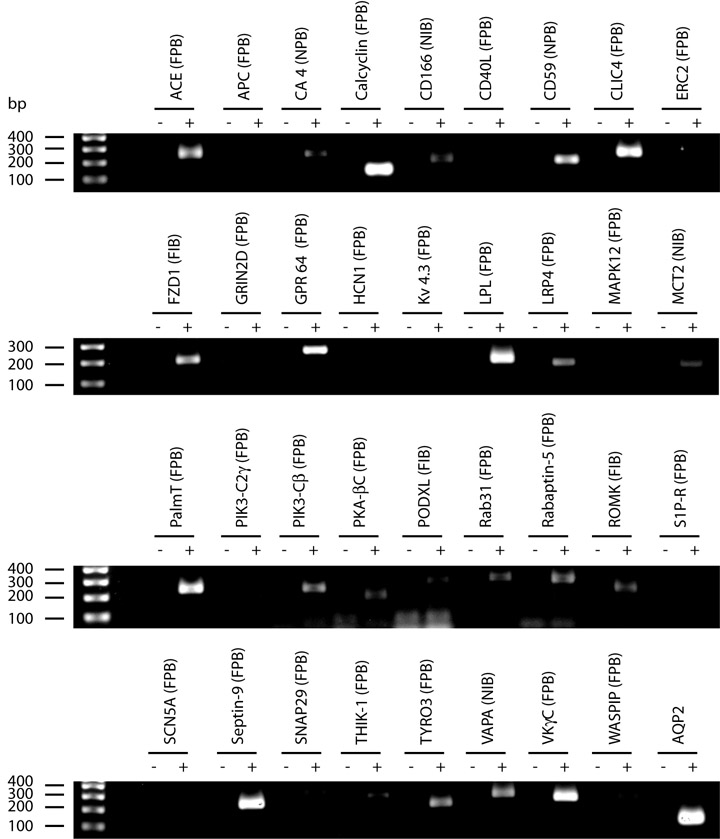
We used biotinylation and streptavidin affinity chromatography to label and enrich proteins from apical and basolateral membranes of rat kidney inner medullary collecting ducts (IMCDs) prior to LC-MS/MS protein identification. To enrich apical membrane proteins and bound peripheral membrane proteins, IMCDs were perfusion-labeled with primary amine-reactive biotinylation reagents at 2 degrees C using a double barreled pipette. The perfusion-biotinylated proteins and proteins bound to them were isolated with CaptAvidin-agarose beads, separated with SDS-PAGE, and sliced into continuous gel pieces for LC-MS/MS protein identification (LTQ, Thermo Electron Corp.). 17 integral and glycosylphosphatidylinositol (GPI)-linked membrane proteins and 44 non-integral membrane proteins were identified. Immunofluorescence confocal microscopy confirmed ACVRL1, H(+)/K(+)-ATPase alpha1, NHE2, and TauT expression in the IMCDs. Basement membrane and basolateral membrane proteins were biotinylated via incubation of IMCD suspensions with biotinylation reagents on ice. 23 integral and GPI-linked membrane proteins and 134 non-integral membrane proteins were identified. Analyses of non-integral membrane proteins preferentially identified in the perfusion-biotinylated and not in the incubation-biotinylated IMCDs revealed protein kinases, scaffold proteins, SNARE proteins, motor proteins, small GTP-binding proteins, and related proteins that may be involved in vasopressin-stimulated AQP2, UT-A1, and ENaC regulation. A World Wide Web-accessible database was constructed of 222 membrane proteins (integral and GPI-linked) from this study and prior studies.
Figures
Similar articles
-
Large-scale quantitative LC-MS/MS analysis of detergent-resistant membrane proteins from rat renal collecting duct.Am J Physiol Cell Physiol. 2008 Sep;295(3):C661-78. doi: 10.1152/ajpcell.90650.2007. Epub 2008 Jul 2. Am J Physiol Cell Physiol. 2008. PMID: 18596208 Free PMC article.
-
High-throughput identification of IMCD proteins using LC-MS/MS.Physiol Genomics. 2006 Apr 13;25(2):263-76. doi: 10.1152/physiolgenomics.00214.2005. Epub 2006 Jan 31. Physiol Genomics. 2006. PMID: 16449382 Free PMC article.
-
Proteomic profiling of nuclei from native renal inner medullary collecting duct cells using LC-MS/MS.Physiol Genomics. 2010 Feb 4;40(3):167-83. doi: 10.1152/physiolgenomics.00148.2009. Epub 2009 Dec 8. Physiol Genomics. 2010. PMID: 19996160 Free PMC article.
-
Phosphorylation of UT-A1 on serine 486 correlates with membrane accumulation and urea transport activity in both rat IMCDs and cultured cells.Am J Physiol Renal Physiol. 2010 Apr;298(4):F935-40. doi: 10.1152/ajprenal.00682.2009. Epub 2010 Jan 13. Am J Physiol Renal Physiol. 2010. PMID: 20071460 Free PMC article.
-
Large scale protein identification in intracellular aquaporin-2 vesicles from renal inner medullary collecting duct.Mol Cell Proteomics. 2005 Aug;4(8):1095-106. doi: 10.1074/mcp.M500049-MCP200. Epub 2005 May 18. Mol Cell Proteomics. 2005. PMID: 15905145 Free PMC article.
Cited by
-
Apicobasal Surfaceome Architecture Encodes for Polarized Epithelial Functionality and Depends on Tumor Suppressor PTEN.Int J Mol Sci. 2022 Dec 19;23(24):16193. doi: 10.3390/ijms232416193. Int J Mol Sci. 2022. PMID: 36555834 Free PMC article.
-
Flow resistance along the rat renal tubule.Am J Physiol Renal Physiol. 2018 Nov 1;315(5):F1398-F1405. doi: 10.1152/ajprenal.00219.2018. Epub 2018 Aug 8. Am J Physiol Renal Physiol. 2018. PMID: 30089029 Free PMC article.
-
Proteomic determination of the lysine acetylome and phosphoproteome in the rat native inner medullary collecting duct.Physiol Genomics. 2018 Sep 1;50(9):669-679. doi: 10.1152/physiolgenomics.00029.2018. Epub 2018 Jun 22. Physiol Genomics. 2018. PMID: 29932826 Free PMC article.
-
Chloride intracellular channel 4 is critical for the epithelial morphogenesis of RPE cells and retinal attachment.Mol Biol Cell. 2010 Sep 1;21(17):3017-28. doi: 10.1091/mbc.E09-10-0907. Epub 2010 Jul 7. Mol Biol Cell. 2010. PMID: 20610659 Free PMC article.
-
Sequence-based searching of custom proteome and transcriptome databases.Physiol Rep. 2018 Sep;6(18):e13846. doi: 10.14814/phy2.13846. Physiol Rep. 2018. PMID: 30230259 Free PMC article.
References
-
- Ecelbarger CA, Terris J, Frindt G, Echevarria M, Marples D, Nielsen S, Knepper MA. Aquaporin-3 water channel localization and regulation in rat kidney. Am J Physiol. 1995;269:F663–F672. - PubMed
-
- Ecelbarger CA, Kim GH, Terris J, Masilamani S, Mitchell C, Reyes I, Verbalis JG, Knepper MA. Vasopressin-mediated regulation of epithelial sodium channel abundance in rat kidney. Am J Physiol. 2000;279:F46–F53. - PubMed
-
- Nicco C, Wittner M, DiStefano A, Jounier S, Bankir L, Bouby N. Chronic exposure to vasopressin upregulates ENaC and sodium transport in the rat renal collecting duct and lung. Hypertension. 2001;38:1143–1149. - PubMed
Publication types
MeSH terms
Substances
Grants and funding
LinkOut - more resources
Full Text Sources
