. 2006 Aug 9;26(32):8310-9.
doi: 10.1523/JNEUROSCI.1779-06.2006.

Integration of visuospatial and effector information during symbolically cued limb movements in monkey lateral intraparietal area

Affiliations

Integration of visuospatial and effector information during symbolically cued limb movements in monkey lateral intraparietal area

Jeff Oristaglio et al. J Neurosci. .

Abstract

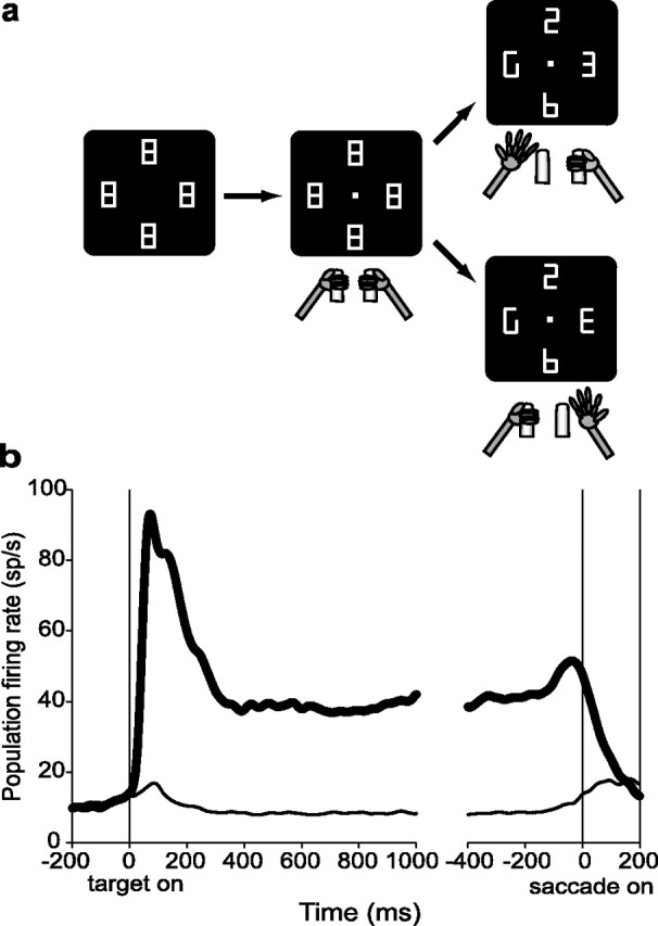
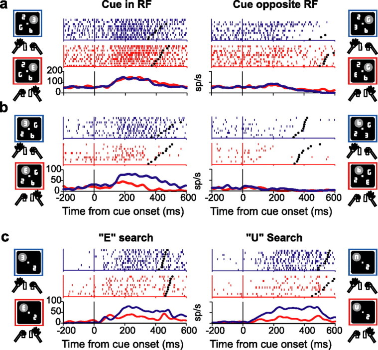
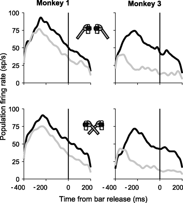
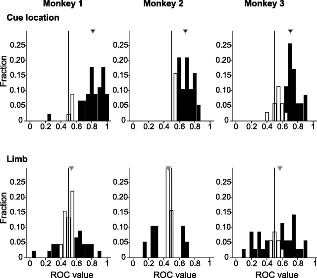
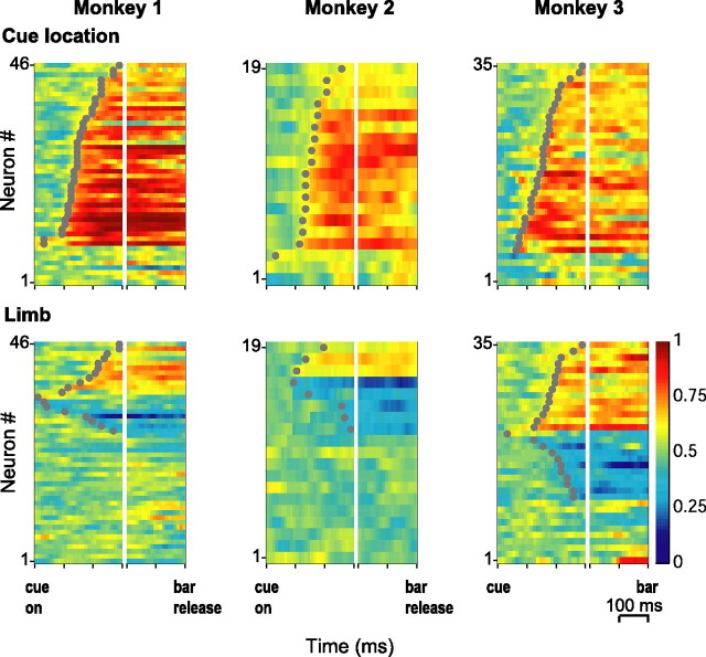
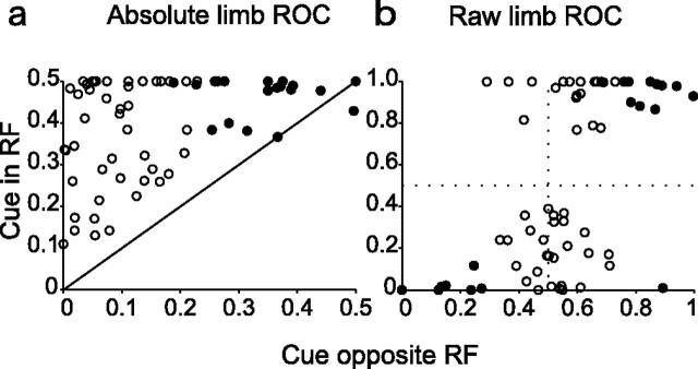
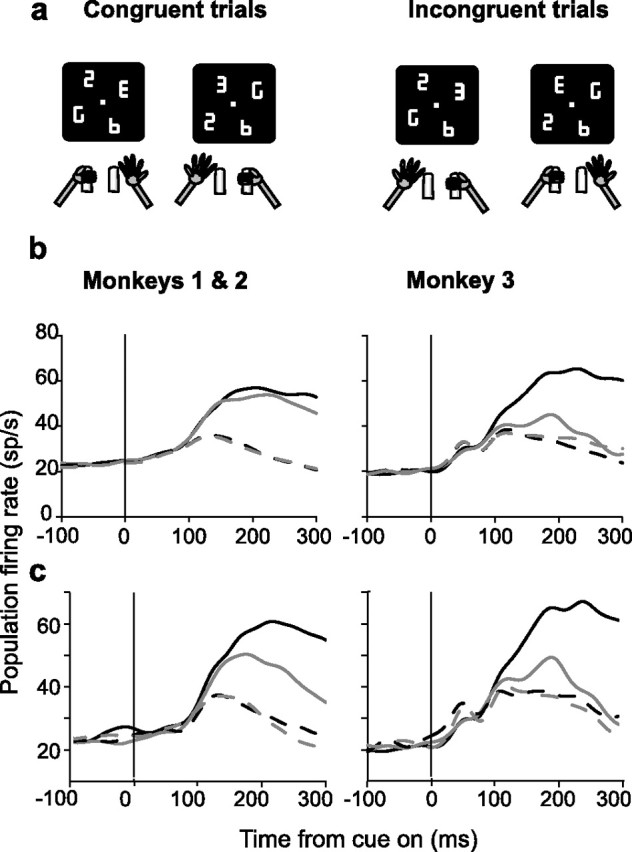
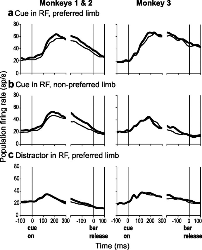
Natural behavior requires close but flexible coordination between attention, defined as selection for perception, and action. In recent years a distributed network including the lateral intraparietal area (LIP) has been implicated in visuospatial selection for attention and rapid eye movements (saccades), but the relation between the attentional and motor functions of this area remains unclear. Here we tested LIP neurons in a task that involved not an ocular but a manual operant response. Monkeys viewed a display containing one cue and several distractors and reported the orientation of the cue (right- or left-facing) by releasing one of two bars grasped, respectively, with the right or left hand. The movement in this task thus was associated with (cued by), but not directed toward, the visual stimulus. A large majority of neurons responded more when the cue rather than when a distractor was in their receptive field, suggesting that they contribute to the attentional selection of the cue. A fraction of these neurons also was modulated by limb release, thus simultaneously encoding cue location and the active limb. The results suggest that the LIP links behaviorally relevant visual information with motor variables relevant for solving a task in a wide range of circumstances involving goal-directed or symbolically cued movements and eye as well as limb movements. A central function of the LIP may be to coordinate visual and motor selection during a wide variety of behaviors.


Figures

Figure 1.
Figure 1.
Behavioral task. a, Each panel illustrates the visual display at different stages of the task. Four placeholders remained stably on the screen throughout the intertrial interval (left). To begin a trial, the monkeys fixated on a central spot and grabbed two bars at stomach level (center). At ∼500 ms after fixation achievement, two line segments were removed from each placeholder, revealing one cue, the letter E, and three distractors (right). Unpredictably and with uniform probabilities, the E appeared at any display location and could be forward-facing (bottom right) or backward-facing (top right). Monkeys received a juice reward if they indicated the orientation of the E by releasing the right hand if the E was forward-facing or the left hand if it was backward-facing. Trials ended with a reward (if correct), extinction of the fixation point, and restoration of the placeholder display. b, Average population activity (81 neurons in monkeys 1 and 3) on the memory saccade task. The thick trace represents activity associated with saccades toward the RF center. The thin trace is the averaged activity for the other three locations tested with the search task for each neuron (90, 180, and −90° from the RF center). Responses are aligned on cue onset (left) and saccade beginning (right).
Figure 2.
Figure 2.
Representative neurons. a, Neuron selective only for cue location. Neural activity is represented in raster plots and averaged spike density histograms from trials in which the cue was either in the center of the RF or at the opposite location and was facing either to the right (red) or to the left (blue). In the raster plots each line is one trial; each tick represents the time of one action potential relative to cue presentation. Spike density histograms were derived by convolving individual spike times with a Gaussian kernel with SD of 15 ms. Activity is aligned on cue presentation (time 0), and black dots represent the bar release. Only correct trials are shown, ordered off-line according to reaction time. Diagrams indicate the location of the cue, the RF (gray oval), and the limb released in each configuration. This neuron responded when the cue was in its RF (top right quadrant) regardless of manual release. b, Neuron selective for cue location and manual release. c, Neuron selective for left-hand release on both E and U search (trials in which the cue was in the RF). Neurons a and b were recorded in monkey 1 and neuron c in monkey 3.
Figure 3.
Figure 3.
Responses in the standard and crossed hand conditions. Population activity from trials in which the cue appeared in the RF is aligned on the time of the bar release for responses with the preferred (black) and nonpreferred (gray) limb. Testing was conducted with an array size of two elements in blocks of trials performed with hands in uncrossed (top) and crossed (bottom) positions.
Figure 4.
Figure 4.
Distribution of ROC indices for cue location (top) and limb (bottom) in the 200 ms before bar release. Filled triangles show the median index, with black denoting a significant difference from 0.5 (p < 0.05; permutation test). For cue location ROC the values above and below 0.5 indicate higher firing rates for, respectively, a cue or a distractor in the RF. For limb ROC the values above and below 0.5 indicate higher firing rates for the right and left limb.
Figure 5.
Figure 5.
ROC indices for cue location and limb. Each color map shows the ROC values in one subject, indicating sensitivity for cue location (top) and limb (bottom). Each row represents one neuron, and each column represents a 10 ms time bin aligned on cue onset (left) and bar release (right). Within each panel, the neurons are sorted by time of onset of significant selectivity (gray dots) and by the direction of this selectivity, with nonselective neurons at the bottom, followed by those with ROC values below 0.5 and above 0.5. For clarity of presentation, the sorting order in the bottom left panel was reversed between neurons with opposite selectivity. Sorting was independent within each panel, and corresponding rows do not indicate the same neurons in the top and bottom panels. The few neurons with very early limb or location selectivity (< 50 ms) most likely reflect preexisting motor bias rather than true accumulation of information within a trial.
Figure 6.
Figure 6.
Limb selectivity as a function of cue location. a, Scatter plots of absolute limb ROC (absolute difference between limb ROC and 0.5) obtained for trials in which the cue was inside or opposite the RF. Filled symbols show neurons with significant selectivity for both cue and distractor in the RF. The diagonal line is the identity line. In monkeys 1–3 16, 6, and 22 neurons showed significant limb effects only when the cue was in the RF, and 5, 3, and 11 neurons showed limb selectivity at both cue locations. b, Same as a but for raw (not absolute) limb ROC. ROC values above and below 0.5 indicate, respectively, preference for the right and left limbs.
Figure 7.
Figure 7.
Congruence. a, Diagrams illustrating congruent and incongruent trials with cues in the right and left hemifield. b, Average population activity from trials in which the cue was either in the RF (solid) or at the opposite location (dashed) and the monkey released the limb congruent or incongruent with the hemifield of the cue (black vs gray). c, Average activity for neurons with significant limb effects from trials in which the cue was either in the RF (solid line) or at the opposite location (dashed line) and the monkey released the preferred or nonpreferred limb for each neuron (black vs gray).
Figure 8.
Figure 8.
Neural activity and reaction time. a, Average population response from trials in which the cue was in the RF, the monkey released the preferred limb of the neurons, and reaction times were either faster (thick) or slower (thin) than the median. b, Same as a, but for release of the nonpreferred limb. c, Average population response trials in which a distractor was in the RF and the animal released the preferred limb with fast (thick) or slow (thin) reaction times.

References

    1. Barash S, Bracewell RM, Fogassi L, Gnadt JW, Andersen RA (1991). Saccade-related activity in the lateral intraparietal area. I. Temporal properties. J Neurophysiol 66:1095–1108. - PubMed
    1. Bisley JW, Goldberg ME (2003). Neuronal activity in the lateral intraparietal area and spatial attention. Science 299:81–86. - PubMed
    1. Calton JL, Dickinson AR, Snyder LH (2002). Non-spatial, motor-specific activation in posterior parietal cortex. Nat Neurosci 5:580–588. - PubMed
    1. Cavanaugh J, Wurtz RH (2004). Subcortical modulation of attention counters change blindness. J Neurosci 24:11236–11243. - PMC - PubMed
    1. Colby CL, Duhamel J-R, Goldberg ME (1996). Visual, presaccadic, and cognitive activation of single neurons in monkey lateral intraparietal area. J Neurophysiol 76:2841–2852. - PubMed

Publication types