. 2006 Aug 9;26(32):8332-8.
doi: 10.1523/JNEUROSCI.1989-06.2006.

Tissue injury regulates serotonin 1D receptor expression: implications for the control of migraine and inflammatory pain

Affiliations

Tissue injury regulates serotonin 1D receptor expression: implications for the control of migraine and inflammatory pain

Andrew H Ahn et al. J Neurosci. .

Abstract

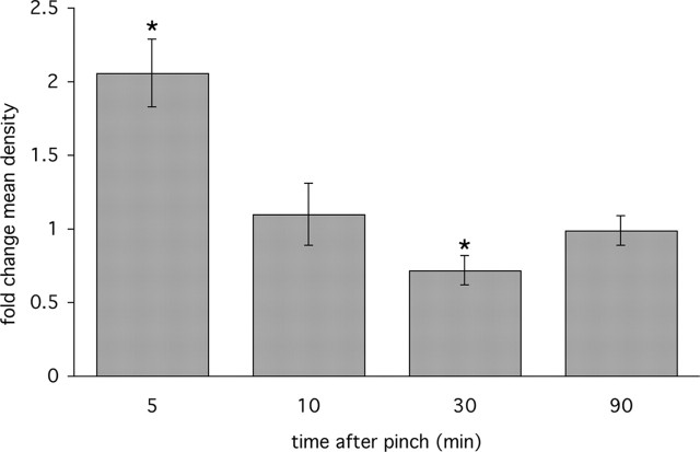
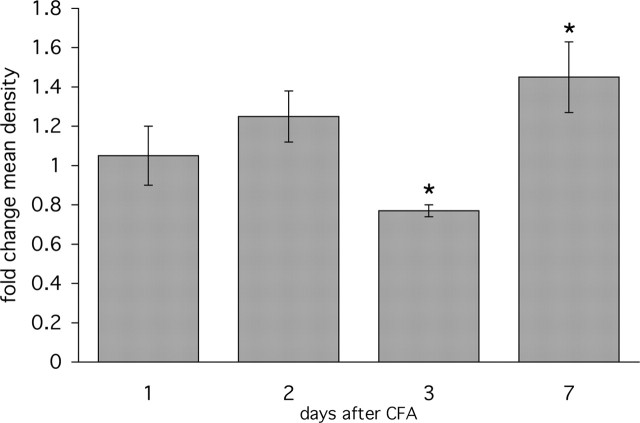
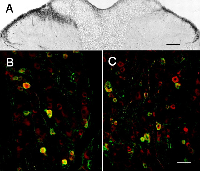
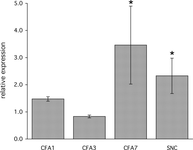
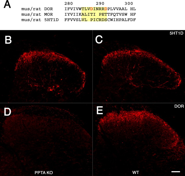
The anti-migraine action of "triptan" drugs involves the activation of serotonin subtype 1D (5-HT1D) receptors expressed on "pain-responsive" trigeminal primary afferents. In the central terminals of these nociceptors, the receptor is concentrated on peptidergic dense core vesicles (DCVs) and is notably absent from the plasma membrane. Based on this arrangement, we hypothesized that in the resting state the receptor is not available for binding by a triptan, but that noxious stimulation of these afferents could trigger vesicular release of DCVs, thus externalizing the receptor. Here we report that within 5 min of an acute mechanical stimulus to the hindpaw of the rat, there is a significant increase of 5-HT1D-immunoreactivity (IR) in the ipsilateral dorsal horn of the spinal cord. We suggest that these rapid immunohistochemical changes reflect redistribution of sequestered receptor to the plasma membrane, where it is more readily detected. We also observed divergent changes in 5-HT1D-IR in inflammatory and nerve-injury models of persistent pain, occurring at least in part through the regulation of 5-HT1D-receptor gene expression. Finally, we found that 5-HT1D-IR is unchanged in the spinal cord dorsal horn of mice with a deletion of the gene encoding the neuropeptide substance P. This result differs from that reported for the partial differential-opioid receptor, which is also sorted to DCVs, but is greatly reduced in preprotachykinin mutant mice. We suggest that a "pain"-triggered regulation of 5-HT1D-receptor expression underlies the effectiveness of triptans for the treatment of migraine. Moreover, the widespread expression of 5-HT1D receptor in somatic nociceptive afferents suggests that triptans could, in certain circumstances, treat pain in nontrigeminal regions of the body.


Figures

Figure 1.
Figure 1.
Dynamic regulation of 5-HT1D-IR in the dorsal horn of the spinal cord after acute mechanical stimulation. L5 lumbar spinal cord staining for 5-HT1D-IR in an animal perfused 5 min (A) and 30 min (B) after mechanical pinch shows increased 5-HT1D-IR at 5 min and reduced staining at 30 min on the stimulated (right) side, compared with the contralateral unstimulated side (control). Scale bar, 100 μm.
Figure 2.
Figure 2.
Quantitative analysis of L5 lumbar spinal cord 5-HT1D-IR in a time series of animals after pinch stimulus of the hindpaw shows the rapid increase of staining at 5 min followed by a reduction in staining at 30 min, and a normalization at 90 min. Fold changes in mean optical density are represented as a ratio of the mean optical density in the medial half of the ipsilateral over the contralateral dorsal horn (*p < 0.05). The levels of 5-HT1D-IR at other spinal cord segments do not differ on the two sides of the cord; the same is true for sections of the L5 segment from untreated animals.
Figure 3.
Figure 3.
Unilateral sensitization of the hindpaw by the inflammatory agent CFA shows dynamic changes in 5-HT1D-IR in the ipsilateral dorsal horn. A, After 3 d, the ipsilateral (right) side of the spinal cord shows reduced 5-HT1D-IR compared with the untreated side (control). B, After 7 d, the level of 5-HT1D-IR is increased ipsilateral to the stimulus. C, The L5 dorsal root ganglia ipsilateral to CFA stimulation at 7 d continues to show significant colocalization of 5-HT1D-IR (red) and substance P (green), whereas (D) 5-HT1D-IR neurons (red) are present in only a small minority of myelinated afferents that immunostain for NF200 (green). Scale bars: (in B) A, B, 100 μm; (in D) C, D, 20 μm.
Figure 4.
Figure 4.
Time course of 5-HT1D-IR after unilateral CFA-induced inflammation. Ipsilateral hindpaw inflammation produces complex changes in 5-HT1D-IR in the medial half of the L5 spinal cord dorsal horn. Levels of 5-HT1D-IR are reduced at 3 d post-CFA, but rise significantly compared with the contralateral side at day 7. *p < 0.05. Error bars are SEM.
Figure 5.
Figure 5.
Differential DRG and spinal cord expression of 5-HT1D-IR after peripheral nerve injury. A, 5-HT1D-IR in the dorsal horn 21 d after complete sciatic nerve transection shows significant reduction of immunoreactivity in the medial portion of the dorsal horn ipsilateral (right) to the injury. Double labeling of L5 dorsal root ganglia for 5-HT1D (red) and CGRP (green) contralateral (B) and ipsilateral (C) to the sciatic nerve transection shows the persistence of receptor expression in peptidergic cell bodies despite its dramatic reduction in their central terminals. Scale bars: A, 100 μm; (in C) B, C, 20 μm.
Figure 6.
Figure 6.
Alteration of 5-HT1D gene expression in chronic models of inflammatory and neuropathic pain. Real-time PCR determination of levels of 5-HT1D mRNA in individual L5 DRG were determined in triplicate and normalized to GAPDH mRNA detected in parallel reactions. Fold changes are expressed as the ratio of levels of cDNA ipsilateral to the injury, compared with the contralateral untreated DRG (see Materials and Methods). Three animals were used for each time point at 1, 3, and 7 d after unilateral intraplantar CFA injection and 21 d after unilateral sciatic nerve cut (SNC). *p < 0.05. Error bars are SEM.
Figure 7.
Figure 7.
The 5-HT1D receptor is sorted to DCVs independently of DOR and SP. A, Alignment of the DOR, MOR, and 5-HT1D receptors in the region of the putative SP-binding domain in the third luminal loop of the DOR, highlighted in yellow. The homologous region of the MOR was used in domain-swapping experiments by Guan et al. (2005) and shown to neither bind SP nor traffic the chimeric DOR to DCVs. This alignment demonstrates a significant divergence of both the MOR and 5-HT1D from key features of the putative binding domain in the DOR. Specifically, the 5-HT1D receptor lacks the aspartate (D) residues at position 288 and 293 (in red), which when mutated greatly diminish interactions between expressed portions of the DOR and SP. The predicted amino acid sequences from mouse and rat are identical for each of the three receptors. 5-HT1D immunofluorescence within the mouse spinal cord dorsal horn of PPT-A mutant mice (B), which lack SP, and in their wild-type littermates (C), is comparable. In contrast, immunostaining of adjacent sections for the DOR shows that it is greatly reduced in PPT-A mutant mice (D) compared with their wild-type littermates (E). Scale bar, 40 μm.

Similar articles

Cited by

References

    1. Abbadie C, Trafton J, Liu H, Mantyh PW, Basbaum AI (1997). Inflammation increases the distribution of dorsal horn neurons that internalize the neurokinin-1 receptor in response to noxious and non-noxious stimulation. J Neurosci 17:8049–8060. - PMC - PubMed
    1. Ahn AH, Basbaum AI (2005). Where do triptans act in the treatment of migraine? Pain 115:1–4. - PMC - PubMed
    1. Antonaci F, Pareja JA, Caminero AB, Sjaastad O (1998). Chronic paroxysmal hemicrania and hemicrania continua: lack of efficacy of sumatriptan. Headache 38:197–200. - PubMed
    1. Bao L, Jin SX, Zhang C, Wang LH, Xu ZZ, Zhang FX, Wang LC, Ning FS, Cai HJ, Guan JS, Xiao HS, Xu ZQ, He C, Hökfelt T, Zhou Z, Zhang X (2003). Activation of delta opioid receptors induces receptor insertion and neuropeptide secretion. Neuron 37:121–133. - PubMed
    1. Bartsch T, Knight YE, Goadsby PJ (2004). Activation of 5-HT(1B/1D) receptor in the periaqueductal gray inhibits nociception. Ann Neurol 56:371–381. - PubMed

Publication types

Substances