. 2006 Aug 9;26(32):8368-76.
doi: 10.1523/JNEUROSCI.0271-06.2006.

Reward-related cortical inputs define a large striatal region in primates that interface with associative cortical connections, providing a substrate for incentive-based learning

Affiliations

Reward-related cortical inputs define a large striatal region in primates that interface with associative cortical connections, providing a substrate for incentive-based learning

Suzanne N Haber et al. J Neurosci. .

Abstract

The anterior cingulate and orbital cortices and the ventral striatum process different aspects of reward evaluation, whereas the dorsolateral prefrontal cortex and the dorsal striatum are involved in cognitive function. Collectively, these areas are critical to decision making. We mapped the striatal area that receives information about reward evaluation. We also explored the extent to which terminals from reward-related cortical areas converge in the striatum with those from cognitive regions. Using three-dimensional-rendered reconstructions of corticostriatal projection fields along with two-dimensional chartings, we demonstrate the reward and cognitive territories in the primate striatum and show the convergence between these cortical inputs. The results show two labeling patterns: a focal projection field that consists of densely distributed terminal patches, and a diffuse projection consisting of clusters of fibers, extending throughout a wide area of the striatum. Together, these projection fields demonstrate a remarkably large, rostral, reward-related striatal territory that reaches into the dorsal striatum. Fibers from different reward-processing and cognitive cortical areas occupy both separate and converging territories. Furthermore, the diffuse projection may serve a separate integrative function by broadly disseminating general cortical activity. These findings show that the rostral striatum is in a unique position to mediate different aspects of incentive learning. Furthermore, areas of convergence may be particularly sensitive to dopamine modulation during decision making and habit formation.


Figures

Figure 1.
Figure 1.
Injection site placements. a, Dorsolateral view of the brain showing the DLPFC injection sites in yellow; b, medial view showing the dACC and vmPFC injection sites in orange and red, respectively; c, ventral view showing the OFC injection sites in dark orange. Note that the injection sites cover a relatively small, restricted part of each functional region. cgs, Cingulate sulcus; cs, central sulcus; iar, inferior arcuate sulcus; lorb, lateral orbital sulcus; morb, medial orbital sulcus; ps, principal sulcus; ros, rostral sulcus; sar, superior arcuate sulcus.
Figure 2.
Figure 2.
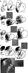
Schematic chartings and microphotographs of labeled fibers after injections into different prefrontal regions. a, vmPFC injection site (area 25); b, dACC injection site (area 24b); c, OFC injection site (area 11); d, DLPFC injection site (area 9/46). The focal projection fields (black area) and the diffuse projections are shown in charts at three anteroposterior levels, rostral to the anterior commissure. e illustrates microphotographs at low and high magnification (1.6×, scale bar, 5 mm; 10×, scale bar, 100 μm) of the dense terminal field charted in d (the most caudal section) with the border of the focal projection. Cd, Caudate nucleus; ic, internal capsule; Pu, putamen nucleus.
Figure 3.
Figure 3.
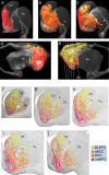
3-D renderings of the combined projection fields from the PFC regions. a–c, Frontal view of the striatum showing inputs from the vmPFC (a), from both dACC and OFC (b), and from all PFC areas, including DLPFC (c); d, e, medial (d) and lateral (e) views showing all PFC inputs. White lines in e indicate the level of sections illustrated in f–j. f–j, Coronal slices through the 3-D model with their corresponding Nissl section: red/purple, Inputs from vmPFC; dark orange, inputs from OFC; light orange, inputs from dACC; yellow, inputs from DLPFC. ac, Anterior commissure; Cd, caudate nucleus; ic, internal capsule; Pu, putamen nucleus.
Figure 4.
Figure 4.
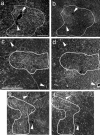
Photomicrographs of adjacent coronal sections of the striatum showing the labeled terminals after dual tracer injections into different PFC areas. a, b, Labeled striatal fibers after injections into dACC and OFC (area 24b, a; area 13, b). Scale bar, 500 μm. c, d, Labeled striatal fibers after injections into vmPFC and OFC (area 14, c; area 13, d). Scale bar, 500 μm. e, f, Labeled striatal fibers after injections into dACC and OFC (area 32, e; area 11, f). The scale bar is shown in c. White outline on a, c, and e indicate the focal projection from areas 24b, 14, and 32, respectively. Note the converging and interdigitation of the labeled clusters. The white arrowheads indicate the correspondent blood vessels in adjacent sections.
Figure 5.
Figure 5.
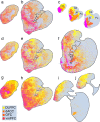
Charts of labeled terminals at different anteroposterior levels of the striatum. a, b, Collective diffuse projections from the ACC and OFC. c, The focal projection fields are represented as colored areas in the small schematics; d–f, interface between ACC/OFC diffuse projections and those from the DLPFC; g–j, diffuse projections are superimposed onto the dense projection showing the interface of the diffuse and focal projections. Cd, Caudate nucleus; ic, internal capsule; Pu, putamen nucleus.
Figure 6.
Figure 6.
After a small PHA-L injection into area 24a, single fibers are found in the dorsal striatum (a), in the area that receives its focal input from area 9m (b). Scale bar, 100 μm.

Similar articles

Cited by

References

    1. Alexander GE, Crutcher MD, DeLong MR (1990). Basal ganglia-thalamocortical circuits: parallel substrates for motor, oculomotor, “prefrontal” and “limbic” functions. Prog Brain Res 85:119–146. - PubMed
    1. Barbas H, Pandya DN (1989). Architecture and intrinsic connections of the prefrontal cortex in the rhesus monkey. J Comp Neurol 286:353–375. - PubMed
    1. Carmichael ST, Price JL (1996). Connectional networks within the orbital and medial prefrontal cortex of macaque monkeys. J Comp Neurol 371:179–207. - PubMed
    1. Charpier S, Mahon S, Deniau JM (1999). In vivo induction of striatal long-term potentiation by low-frequency stimulation of the cerebral cortex. Neuroscience 91:1209–1222. - PubMed
    1. Corlett PR, Aitken MR, Dickinson A, Shanks DR, Honey GD, Honey RA, Robbins TW, Bullmore ET, Fletcher PC (2004). Prediction error during retrospective revaluation of causal associations in humans: fMRI evidence in favor of an associative model of learning. Neuron 44:877–888. - PubMed

Publication types