. 2006 Aug 9;26(32):8398-408.
doi: 10.1523/JNEUROSCI.0618-06.2006.

Calcium activation of the LMO4 transcription complex and its role in the patterning of thalamocortical connections

Affiliations

Calcium activation of the LMO4 transcription complex and its role in the patterning of thalamocortical connections

Amir H Kashani et al. J Neurosci. .

Abstract

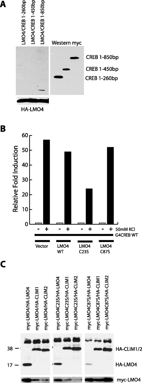
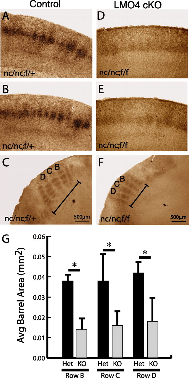
Lasting changes in neuronal connectivity require calcium-dependent gene expression. Here we report the identification of LIM domain-only 4 (LMO4) as a mediator of calcium-dependent transcription in cortical neurons. Calcium influx via voltage-sensitive calcium channels and NMDA receptors contributes to synaptically induced LMO4-mediated transactivation. LMO4-mediated transcription is dependent on signaling via calcium/calmodulin-dependent protein (CaM) kinase IV and microtubule-associated protein (MAP) kinase downstream of synaptic stimulation. Coimmunoprecipitation experiments indicate that LMO4 can form a complex with cAMP response element-binding protein (CREB) and can interact with cofactor of LIM homeodomain protein 1 (CLIM1) and CLIM2. To evaluate the role of LMO4 in vivo, we examined the consequences of conditional loss of lmo4 in the forebrain, using the Cre-Lox gene-targeting strategy. The organization of the barrel field in somatosensory cortex is disrupted in mice in which lmo4 is deleted conditionally in the cortex. Specifically, in contrast to controls, thalamocortical afferents in conditional lmo4 null mice fail to segregate into distinct barrel-specific domains. These observations identify LMO4 as a calcium-dependent transactivator that plays a key role in patterning thalamocortical connections during development.


Figures

Figure 1.
Figure 1.
Depolarization-induced activation of LMO4-mediated transcription and expression pattern of LMO4. A, B, E18 dissociated cortical neurons at 5 DIV immunostained for CAT after being transfected with GAL4-LMO4 and UAS-CAT and either left untreated or stimulated with 50 mm KCl. C, Northern blot of RNA isolated from cortices at different developmental ages hybridized with probes to LMO4 and CLIM2. Then, the same blot was stripped and hybridized with a glyceraldehyde-3-phosphate dehydrogenase (GAPDH) probe for loading control. D–F, Distribution of LMO4 in the developing cortex detected by immunofluorescence with the use of goat anti-LMO4 (green) and mouse anti-MAP2 (red) antibodies on coronal slices of P15 mouse cortex. G–I, Subcellular localization of LMO4 (green) in E18 dissociated cortical cultures counterstained with a nuclear Hoechst dye (red).
Figure 2.
Figure 2.
Characterization of LMO4-mediated transcription. E18 dissociated cortical neurons were transfected with the indicated constructs at 3 DIV and treated as indicated at 5 DIV. A, Relative CAT activity in neurons transfected with GAL4-DBD, GAL4-CREB, or GAL4-LMO4 and UAS-CAT and stimulated with 50 mm KCl. B, Relative CAT activity of neurons transfected with GAL4-LHX2 and UAS-CAT and stimulated with 50 mm KCl. C, Relative CAT activity of neurons transfected with GAL4-LMO4 and UAS-CAT and pretreated with pharmacologic inhibitors as indicated, followed by 50 mm KCl stimulation. D, Relative CAT activity of neurons transfected with wild-type GAL4-LMO4 or constructs with cysteine-to-serine point mutations at either cysteine 23 or 87, as indicated, and stimulated with 50 mm KCl. Asterisks indicate significance at p < 0.05. Error bars represent ± SEM.
Figure 3.
Figure 3.
Signaling pathways involved in calcium activation of LMO4-mediated transcription. A, Relative CAT activity in E18 dissociated cortical neurons transfected with GAL4-LMO4 and either wild-type or constitutively active forms of CaMK II (CKII), CaMK IV (CKIV), or MEK at 3 DIV and assayed for CAT activity at 5 DIV. B, D, F, Relative CAT activity in E18 dissociated cortical neurons transfected with GAL4-LMO4 at 3 DIV and pretreated with pharmacologic inhibitors, followed by 50 mm KCl stimulation at 5 DIV. C, E, Relative CAT activity in E18 dissociated cortical neurons transfected with GAL4-LMO4 and UAS-CAT, together with indicated constructs, at 3 DIV and stimulated as indicated at 5 DIV. Asterisks indicate significance at p < 0.05. Error bars represent ± SEM.
Figure 4.
Figure 4.
Synaptic stimulation induces LMO4-mediated transcription via NMDAR and L-VSCC activation. A, Relative CAT activity in E18 dissociated cortical neurons transfected with GAL4-LMO4 and UAS-CAT at 3 DIV and stimulated with either 50 mm KCl or 10 μm glutamate at 5 DIV, as indicated. B, C, Relative CAT activity in E18 cortical cultures transfected with GAL4-LMO4 and UAS-CAT at 3 DIV and treated as indicated at 12–14 DIV. Asterisks indicate significance at p < 0.05. Error bars represent ± SEM.
Figure 5.
Figure 5.
LMO4 interacts with CREB, CLIM1, and CLIM2. A, Coimmunoprecipitation of HA-tagged LMO4 with myc-tagged CREB deletion constructs in 293T-cells. Myc immunoprecipitations followed by anti-HA Western blots show the interaction of LMO4 with CREB base pairs 1–850, but not with CREB deletion constructs containing base pairs 1–260 and 1–450 (top left). Equivalent transfection of HA-LMO4 in all plates is indicated by the HA Western blot on whole-cell lysates (bottom left). Equivalent expression of CREB constructs is indicated by the myc-Western blot (bottom right). B, Relative CAT activity in E18 neurons transfected with GAL4-CREB and UAS-CAT, together with indicated constructs, at 3 DIV and stimulated at 5 DIV. C, Coimmunoprecipitation assays in 293T-cells transfected with myc-tagged LMO4, together with HA-LMO4, HA-CLIM1, or HA-CLIM2. LMO4 can interact with LMO4 as well as with both CLIM1 and CLIM2. Mutation of cysteine 87 to serine [LMO4(C87S)] in the LMO4 protein prevents LMO4 dimerization. LMO4(C23S) and LMO4(C87S) indicate constructs in which the conserved cysteine at position 23 or 87, respectively, was mutated to a serine.
Figure 6.
Figure 6.
Targeted deletion of LMO4 in cortex. A, Cre-recombinase activity in the whole brain of floxed LacZ reporter mice crossed with nexCre transgenics. Recombination is seen in the cortex (cx) and hippocampus (hip). la, Lateral amygdaloid nucleus; pir, piriform cortex. B, High magnification of LacZ expression in the cortex after nexCre-mediated recombination. C, D, LMO4 expression (green) and MAP2 expression (red) in P15 cortex in control mice (C; n = 3) and LMO4 conditional knock-out mice (D; n = 3) detected by immunofluorescence. Genotypes are indicated in each panel (nc, nexCre; f, floxed lmo4). E, F, Immunofluorescence for neuronal (green) and GABAergic (red) markers in cortices of control (E) and LMO4 conditional knock-outs (F). G, Western blot detection of LMO4 protein from isolated cortices of control, lmo4 conditional heterozygous, and lmo4 conditional knock-out animals. H, Genotyping PCR from sample litter of mice bearing various lmo4 alleles (top band, 265 nt floxed allele; bottom band, 180 nt wild-type allele) and nexCre alleles (top band, 770 nt wild-type allele; bottom band, 520 nt Cre-recombination allele). The left panel represents PCR results from two lmo4 conditional knock-outs (nc/nc; f/f). The right panel shows PCR results from heterozygous and wild-type animals.
Figure 7.
Figure 7.
Mice with a conditional deletion of LMO4 have abnormal development of somatosensory barrels. Shown is cytochrome oxidase staining of coronal and tangential sections from control (A, B, E, F; n = 15) and lmo4 conditional knock-out (cKO) cortices (C, D, G, H; n = 9) at P15. Barrels consist of denser patches of staining interspersed with lighter areas (arrows in A and B). Control animals have clearly defined barrel patterns in both coronal and tangential planes, whereas conditional lmo4 knock-outs have either no barrels or very small patches of staining. Genotypes of animals are indicated in the bottom left corner of each panel. Similar results were seen in nine conditional lmo4 knock-out mice and 15 control mice between P15 and P30, indicating that this is not attributable to a developmental delay.
Figure 8.
Figure 8.
Segregation of thalamocortical afferents is disrupted in conditional LMO4 null mice. P7 mice cortices were fixed, sectioned, and stained for 5-HT immunohistochemistry. Coronal (A, B) and tangential (C) sections of control mouse (genotype nc/nc;f/+) reveal normal patterning of thalamocortical afferents as revealed by 5-HT immunohistochemistry. Coronal (D, E) and tangential (F) sections of conditional LMO4 null mice (genotype nc/nc;f/f) reveal poor segregation of thalamocortical afferents with small barrels, poorly defined boundaries, and overall decreased cross-sectional area (G; labeled in C and F as rows B, C, D) compared with controls. Asterisks indicate significance at p < 0.05 by paired Student's t test. Error bars represent ± SEM.

References

    1. Ahn S, Riccio A, Ginty DD (2000). Spatial considerations for stimulus-dependent transcription in neurons. Annu Rev Physiol 62:803–823. - PubMed
    1. Aizawa H, Hu SC, Bobb K, Balakrishnan K, Ince G, Gurevich I, Cowan M, Ghosh A (2004). Dendrite development regulated by CREST, a calcium-regulated transcriptional activator. Science 303:197–202. - PubMed
    1. Bading H, Ginty DD, Greenberg ME (1993). Regulation of gene expression in hippocampal neurons by distinct calcium signaling pathways. Science 260:181–186. - PubMed
    1. Boehm T, Baer R, Lavenir I, Forster A, Waters JJ, Nacheva E, Rabbitts TH (1988). The mechanism of chromosomal translocation t(11;14) involving the T-cell receptor Cδ locus on human chromosome 14q11 and a transcribed region of chromosome 11p15. EMBO J 7:385–394. - PMC - PubMed
    1. Bulchand S, Subramanian L, Tole S (2003). Dynamic spatiotemporal expression of LIM genes and cofactors in the embryonic and postnatal cerebral cortex. Dev Dyn 226:460–469. - PubMed

Publication types

MeSH terms