Photoaffinity labeling combined with mass spectrometric approaches as a tool for structural proteomics
- PMID: 16901199
- PMCID: PMC2266983
- DOI: 10.1586/14789450.3.4.399
Photoaffinity labeling combined with mass spectrometric approaches as a tool for structural proteomics
Abstract
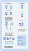
Protein chemistry, such as crosslinking and photoaffinity labeling, in combination with modern mass spectrometric techniques, can provide information regarding protein-protein interactions beyond that normally obtained from protein identification and characterization studies. While protein crosslinking can make tertiary and quaternary protein structure information available, photoaffinity labeling can be used to obtain structural data about ligand-protein interaction sites, such as oligonucleotide-protein, drug-protein and protein-protein interaction. In this article, we describe mass spectrometry-based photoaffinity labeling methodologies currently used and discuss their current limitations. We also discuss their potential as a common approach to structural proteomics for providing 3D information regarding the binding region, which ultimately will be used for molecular modeling and structure-based drug design.
Figures


Similar articles
-
Mass spectrometry-based structural proteomics.Eur J Mass Spectrom (Chichester). 2012;18(2):251-67. doi: 10.1255/ejms.1178. Eur J Mass Spectrom (Chichester). 2012. PMID: 22641729 Review.
-
Nonspecific protein labeling of photoaffinity linkers correlates with their molecular shapes in living cells.Chem Commun (Camb). 2016 Apr 30;52(34):5828-31. doi: 10.1039/c6cc01426g. Epub 2016 Apr 4. Chem Commun (Camb). 2016. PMID: 27043101
-
Photoaffinity labeling and its application in structural biology.Biochemistry (Mosc). 2007 Jan;72(1):1-20. doi: 10.1134/s0006297907010014. Biochemistry (Mosc). 2007. PMID: 17309432 Review.
-
Clickable photoaffinity ligands for the human serotonin transporter based on the selective serotonin reuptake inhibitor (S)-citalopram.Bioorg Med Chem Lett. 2018 Nov 15;28(21):3431-3435. doi: 10.1016/j.bmcl.2018.09.029. Epub 2018 Sep 22. Bioorg Med Chem Lett. 2018. PMID: 30266542 Free PMC article.
-
Identification of alcohol-binding site(s) in proteins using diazirine-based photoaffinity labeling and mass spectrometry.Chem Biol Drug Des. 2019 Jun;93(6):1158-1165. doi: 10.1111/cbdd.13403. Epub 2018 Oct 22. Chem Biol Drug Des. 2019. PMID: 30346111 Free PMC article.
Cited by
-
Photoaffinity labeling of high affinity nicotinic acid adenine dinucleotide phosphate (NAADP)-binding proteins in sea urchin egg.J Biol Chem. 2012 Jan 20;287(4):2308-15. doi: 10.1074/jbc.M111.306563. Epub 2011 Nov 23. J Biol Chem. 2012. PMID: 22117077 Free PMC article.
-
Mass spectrometric approaches to study protein structure and interactions in lyophilized powders.J Vis Exp. 2015 Apr 14;(98):52503. doi: 10.3791/52503. J Vis Exp. 2015. PMID: 25938927 Free PMC article.
-
Probing the conformation of the ISWI ATPase domain with genetically encoded photoreactive crosslinkers and mass spectrometry.Mol Cell Proteomics. 2012 Apr;11(4):M111.012088. doi: 10.1074/mcp.M111.012088. Epub 2011 Dec 13. Mol Cell Proteomics. 2012. PMID: 22167269 Free PMC article.
-
Photocrosslinking approaches to interactome mapping.Curr Opin Chem Biol. 2013 Feb;17(1):90-101. doi: 10.1016/j.cbpa.2012.10.034. Epub 2012 Nov 10. Curr Opin Chem Biol. 2013. PMID: 23149092 Free PMC article. Review.
-
Design, synthesis and evaluation of photoactivatable derivatives of microtubule (MT)-active [1,2,4]triazolo[1,5-a]pyrimidines.Bioorg Med Chem Lett. 2018 Jul 1;28(12):2180-2183. doi: 10.1016/j.bmcl.2018.05.010. Epub 2018 May 5. Bioorg Med Chem Lett. 2018. PMID: 29764743 Free PMC article.
References
-
-
Sing A, Thornton ER, Westheimer FH. The photolysis of diazoacetylchymotrypsin. J. Biol. Chem. 1962;237(9):PC3006–PC3008. • Earliest report of the use of photoaffinity chemistry in biochemistry.
-
-
- Fleming SA. Chemical reagents in photoaffinity labeling. Tetrahedron. 1995;51(46):12479–12520.
-
- Brunner J. New photolabeling and crosslinking methods. Ann. Rev. Biochem. 1993;62:483–514. - PubMed
-
-
Dormán G, Prestwich GD. Using photolabile ligands in drug discovery and development. Trends Biotechnol. 2000;18:64–77. •• Focuses on how photoaffinity labeling techniques are used in drug discovery, but does not delve deeply into mass spectrometry
-
-
-
Weber PJ, Beck-Sickinger AG. Comparison of the photochemical behavior of four different photo-activatable probes. J. Peptide Res. 1997;49(5):375–383. • Useful information for those researchers contemplating the use of photoaffinity techniques.
-
Publication types
MeSH terms
Substances
Grants and funding
LinkOut - more resources
Full Text Sources
Other Literature Sources
