Cytodiagnosis of multiple myeloma presenting as orbital involvement: a case report
- PMID: 16901345
- PMCID: PMC1564147
- DOI: 10.1186/1742-6413-3-19
Cytodiagnosis of multiple myeloma presenting as orbital involvement: a case report
Abstract
Background: Plasma cell neoplasms represent autonomous proliferations of plasma cells and can manifest as diffuse myeloma with systemic involvement (plasma cell myeloma or multiple myeloma), monoclonal gammopathy of undetermined significance (MGUS), or as variants of plasma cell myeloma such as indolent myeloma, smoldering myeloma, osteosclerotic myeloma, plasma cell leukaemia and non-secretory myeloma. Localized neoplastic proliferation of plasma cells presents as solitary plasmacytoma of bone or extramedullary plasmacytoma. Involvement of orbit can occur as a solitary plasmacytoma, or as part of systemic involvement in multiple myeloma, the clinical outcome being significantly worse in the latter setting. Orbital involvement in multiple myeloma is very rare with less than 50 cases reported in the literature. Early cytological diagnosis of such lesions is vital for timely institution of appropriate therapy. As far as we are aware only six previous cases of cytological diagnosis of multiple myeloma involving the orbit are on record.
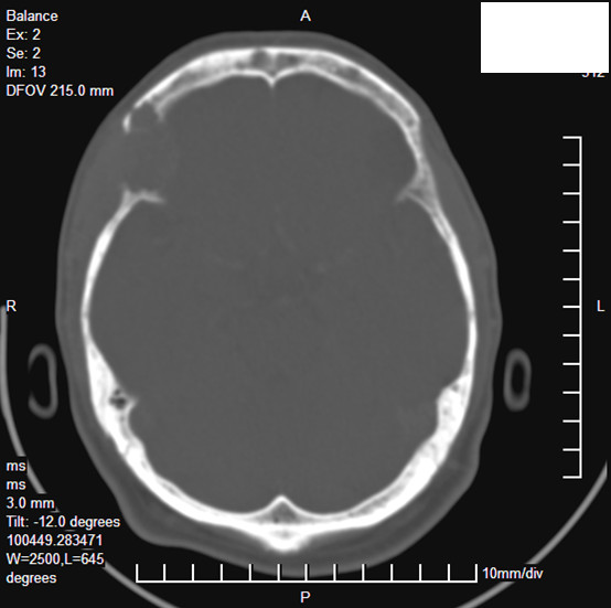
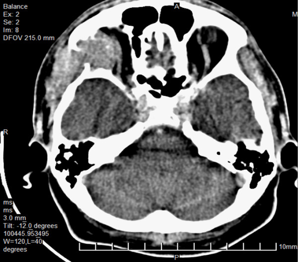
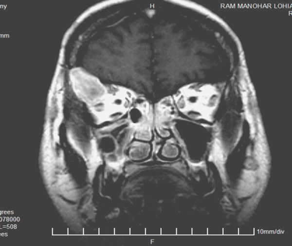
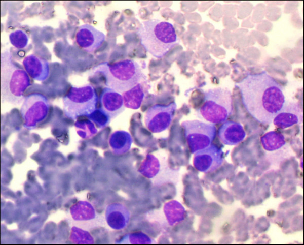
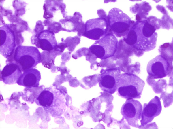
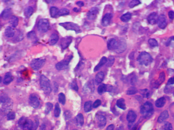
Case presentation: A 37 year old male presented with low grade fever showing evening rise, headache, diplopia and swelling in the right periorbital and temporal region. Imaging studies revealed destructive lesion of sphenoid, frontal bone and zygomatic arch with soft tissue component extending to infratemporal fossa and orbit. A fine needle aspirate from the temporal region swelling showed features of a plasmacytoma, and subsequent workup confirmed the presence of systemic disease. A final diagnosis of multiple myeloma with orbital involvement at presentation was made.
Conclusion: Present case describes the extremely rare presentation of multiple myeloma with orbital involvement and highlights the utility of cytology in such lesions. Fine needle aspiration diagnosis of plasmacytoma at extramedullary sites offers an opportunity for non-invasive verification of systemic involvement, and thus plays a major role in early diagnosis and management of these patients.
Figures
Similar articles
-
Plasma cell tumours: cytomorphological features in a series of 12 cases diagnosed on fine needle aspiration cytology.Cytopathology. 2010 Jun;21(3):186-90. doi: 10.1111/j.1365-2303.2009.0641.x. Epub 2009 Mar 17. Cytopathology. 2010. PMID: 19416310
-
Osteosclerotic plasmacytoma of maxillary bone (orbital floor).J Laryngol Otol. 1984 Sep;98(9):929-38. doi: 10.1017/s0022215100147735. J Laryngol Otol. 1984. PMID: 6481230
-
A rare differential diagnosis for cause of proptosis: skull plasmacytoma.Neuroradiol J. 2012 Jul;25(3):374-8. doi: 10.1177/197140091202500315. Epub 2012 Jun 26. Neuroradiol J. 2012. PMID: 24028993
-
Primary extramedullary plasmacytoma with diffuse lymph node involvement: a case report and review of the literature.J Med Case Rep. 2019 May 22;13(1):153. doi: 10.1186/s13256-019-2087-7. J Med Case Rep. 2019. PMID: 31113466 Free PMC article. Review.
-
Diffuse skeletal muscle extramedullary plasmacytomas: a rare case and review of the literature.Skeletal Radiol. 2020 Dec;49(12):2087-2093. doi: 10.1007/s00256-020-03514-9. Epub 2020 Jun 19. Skeletal Radiol. 2020. PMID: 32556470 Review.
Cited by
-
Bilateral Proptosis in a Case of Recurring Multiple Myeloma: Uncommon Orbital Presentation of Plasmacytoma.Int Med Case Rep J. 2020 Jul 27;13:297-301. doi: 10.2147/IMCRJ.S260472. eCollection 2020. Int Med Case Rep J. 2020. PMID: 32884366 Free PMC article.
-
Exploring the ocular involvement in multiple myeloma: a comprehensive review of 70-year clinical studies.Int Ophthalmol. 2025 Mar 14;45(1):89. doi: 10.1007/s10792-025-03467-9. Int Ophthalmol. 2025. PMID: 40085267 Review.
-
Cytodiagnosis of multiple myeloma presenting as chest wall swellings.J Cytol. 2012 Apr;29(2):135-6. doi: 10.4103/0970-9371.97158. J Cytol. 2012. PMID: 22787296 Free PMC article. No abstract available.
-
Frontal skull craniotomy combined with moderate-dose radiotherapy effectively ameliorate a rare case of non-secretory, multiple myeloma with orbital involvement.World J Surg Oncol. 2009 Nov 12;7:86. doi: 10.1186/1477-7819-7-86. World J Surg Oncol. 2009. PMID: 19909529 Free PMC article.
-
CytoJournal's move to the new platform: More on financial model to the support open-access charter in cytopathology, publication quality indicators, and other issues.Cytojournal. 2008 Dec 16;5:15. doi: 10.4103/1742-6413.44572. Cytojournal. 2008. PMID: 19495401 Free PMC article. No abstract available.
References
-
- Dispenzieri A, Lacy MQ, Greipp PR. In: Multiple Myeloma: In 'Wintrobe's Clinical Hematology'. 11. Greer JP, Foerster J, Lukens JN, Rodgers GM, Paraskevas F, Glader B, editor. Philadelphia: Lippincott Williams & Wilkins; 2004. pp. 2583–2636.
-
- SEER Cancer statistics review 1975–2003 http://seer.cancer.gov/csr/1975_2003/results_merged/sect_18_myeloma.pdf
-
- Howling SJ, Tighe J, Patterson K, Shaw P. Case report: The CT features of orbital multiple myeloma. Clin Radiol. 1998;53 3-4-305. - PubMed
-
- Fay AM, Lieb ML, Fountain KS. Multiple myeloma involving the orbit. Ophthalmic Plast Reconstr Surg. 1998;14:67–71. - PubMed
-
- Rodman HL, Font RL. Orbital involvement in multiple myeloma: a review of literature and report of three cases. Arch Ophthalmol. 1972;87:30–35. - PubMed
LinkOut - more resources
Full Text Sources
Molecular Biology Databases
Research Materials