. 2006 Oct 6;281(40):29703-10.
doi: 10.1074/jbc.M607351200. Epub 2006 Aug 10.

The protein kinase C delta catalytic fragment targets Mcl-1 for degradation to trigger apoptosis

Affiliations

The protein kinase C delta catalytic fragment targets Mcl-1 for degradation to trigger apoptosis

Leonid A Sitailo et al. J Biol Chem. .

Abstract

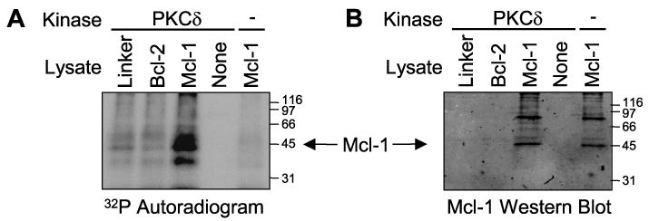
Proteolytic cleavage and subsequent activation of protein kinase C (PKC) delta is required for apoptosis induced by a variety of genotoxic agent, including UV radiation. In addition, overexpression of the constitutively active PKCdelta catalytic fragment (PKCdelta-cat) is sufficient to trigger Bax activation, cytochrome c release, and apoptosis. While PKCdelta is a key apoptotic effector, the downstream target(s) responsible for the mitochondrial apoptotic cascade are not known. We found that expression of the active PKCdelta-cat in HaCaT cells triggers a reduction in the anti-apoptotic protein Mcl-1, similar to UV radiation. The down-regulation of Mcl-1 induced by PKCdelta-cat was not at the mRNA level but was due to decreased protein half-life. Overexpression of Mcl-1 protected HaCaT cells from both UV and PKCdelta-cat-induced apoptosis and blocked the release of cytochrome c from the mitochondria, indicating that Mcl-1 down-regulation was required for apoptosis signaling. Indeed, down-regulation of Mcl-1 with siRNA slightly increased the basal apoptotic rate of HaCaT cells and dramatically sensitized them to UV or PKCdelta-cat-induced apoptosis. HaCaT cells with down-regulated Mcl-1 had higher activated Bax protein, as measured by Bax cross-linking, indicating that Mcl-1 down-regulation is sufficient for Bax activation. Finally, recombinant PKCdelta could phosphorylate Mcl-1 in vitro, identifying Mcl-1 as a direct target for PKCdelta. Overall our results identify Mcl-1 as an important target for PKCdelta-cat that can mediate its pro-apoptotic effects on mitochondria to amplify the apoptotic signaling induced by a wide range of apoptotic stimuli.


Figures

FIGURE 1
FIGURE 1. PKCδ-cat and UV trigger loss of Mcl-1 in HaCaT cells
A, HaCaT cells were infected with either control Linker virus (L) or PKCδ-ER virus and treated with 100 nm Tam for 3 days to activate PKCδ-ER or exposed to UV (30 mJ/cm2, 18 h) as indicated. Western blot analysis for Mcl-1 protein was performed, and the percentage of Mcl-1 relative to Linker virus control is shown. Western blot for β-actin confirmed equal protein loading. B, HaCaT cells were infected with either control Linker virus (L) or PKCδ-ER virus (PKCδ) and treated with 100 nm Tam as indicated for 3 days or exposed to UV (30 mJ/cm2, 18 h). Western blot and quantitation using two different antibodies are shown. The right panel used sc-819 (Santa Cruz Biotechnology), and the right panel used monoclonal antibody 4602 (clone RC13, Chemicon). C, quantitative analysis of Mcl-1 protein level measured from six independent Western blots experiments as described for A. The Mcl-1 levels in Linker-infected cells were normalized to 100%. Error bars represent standard deviation.
FIGURE 2
FIGURE 2. PKCδ-cat and UV reduce Mcl-1 protein half-life in HaCaT cells
A, HaCaT cells were infected with either Linker, PKCδ-ER, or the catalytically inactive PKCδ(K378A)-ER virus and treated with Tam to activate PKCδ-ER or exposed to UV as indicated. Three days after virus infection or immediately after UV exposure, protein synthesis was blocked with 10 μg/ml of cyclohexamide (CHX), and cells were harvested at different time points (0, 0.5, 1, 2, 4, and 6 h). Western blots for Mcl-1 were performed and the intensity of Mcl-1 protein bands determined and normalized to β-actin. The half-life of Mcl-1 protein was estimated as described under “Materials and Methods.” B, Mcl-1 protein half-life was determined as described for A, and data averaged from three experiments are shown. Error bars represent standard deviation. The asterisks indicate significant differences between the treated groups and the Linker + Tam group (p < 0.05). C, the half-life of Mcl-1 protein in HeLa cells was determined as described for A. Cells were treated with CHX alone, exposed to UV, or exposed to UV and treated with CHX and the half-life of Mcl-1 calculated from Mcl-1 Western blots. Note that in HeLa cells Mcl-1 has a short half-life (∼40 min) that is not affected by UV.
FIGURE 3
FIGURE 3. PKCδ-cat does not reduce Mcl-1 mRNA levels
HaCaT cells were infected with Linker, PKCδ-ER, or PKCδ(K378A)-ER viruses and treated with Tam to activate PKCδ-ER or exposed to UV (30 mJ/cm2) as indicated. RNA was isolated 3 days after viral infection or 18 h after UV exposure and an RNase protection assay performed. Hybridization to L32 and glyceraldehyde-3-phosphate dehydrogenase (GAPDH) was used as control to ensure equal levels of input RNA. Note that Mcl-1 mRNA levels were not decreased by PKCδ-cat (PKCδ-ER+Tam).
FIGURE 4
FIGURE 4. Mcl-1 protects from PKCδcat and UV apoptosis
A, HaCaT cells were infected with either control Linker (L) or Mcl-1 (M) viruses and Mcl-1 protein levels determined by Western blotting. Note the overexpression of Mcl-1L and Mcl-1S proteins in Mcl-1-infected cells. B, HaCaT cells were infected with the control Linker or Mcl-1 retroviruses in the morning and in the afternoon re-infected with PKCδ-ER as indicated. The next day cells were treated with Tam for 3 days and phase contrast pictures taken. The cells irradiated with UV (30 mJ/cm2) were photographed after 18 h. Note that the morphological cell death in the PKCδ-ER+Tam and UV-irradiated cells was partially inhibited by Mcl-1. C, HaCaT cells were infected with Linker or Mcl-1 virus and then re-infected with either PKCδ-ER or PKCδ(K378A)-ER viruses and treated with Tam to activate PKCδ-ER or irradiated with UV as indicated. Apoptosis was assayed measured by Annexin V/propidium iodide staining. Note the significant reduction of PKCδ-ER and UV apoptosis by Mcl-1. The catalytically inactive PKCδ(K378A)-ER virus did not induce apoptosis. Data shown are averaged from three independent experiments with error bars representing the standard deviation.
FIGURE 5
FIGURE 5. Mcl-1 blocks PKCδ-cat-induced cytochrome c release
HaCaT cells were infected with Linker, PKCδ-ER, and/or Mcl-1 viruses, treated with Tam to activate PKCδ-ER, or exposed to UV as indicated. The general caspase inhibitor Z-VAD was added at 10 μg/ml as indicated. Cytosolic fractions were analyzed by Western blotting for release of cytochrome c from the mitochondria. Note that PKCδ-cat and UV triggered cytochrome c release that was inhibited by Mcl-1. Z-VAD did not prevent cytochrome c release. A Western blot for β-actin was done as a control of protein loading.
FIGURE 6
FIGURE 6. Suppression of Mcl-1 with siRNA sensitizes HaCaT cells to apoptosis
A, HaCaT cells were infected with control (C) or Mcl-1 siRNA retroviruses and cultured in the presence of 1 μg/ml puromycin for 2 days. Western blots for Mcl-1 and β-actin were performed. Data from three independent experiments are shown, with the percent of Mcl-1 in each experiment normalized to the control. B, HaCaT cells infected with either control or Mcl-1 siRNA viruses were selected with puromycin and then re-infected with PKCδ-cat virus or exposed to UV as indicated. Three days after the PKCδ-cat infection or 18 h after UV irradiation, apoptosis was assayed by Annexin V/propidium iodide staining. Each condition in the experiment was performed in triplicate, with error bars denoting standard deviation. C, HaCaT cells infected with control or Mcl-1 siRNA viruses were selected with puromycin and re-infected with PKCδ-ER virus as indicated. PKCδ activity was induced by addition of Tam to the media of PKCδ-ER-infected cells. Three days after PKCδ-ER infection or 18 h after UV irradiation, the heavy membrane fraction was isolated and a Western blot for Bax performed. Total Bax levels were quantified by densitometry and are indicated at the bottom of the Western blot. Note the increase in total Bax levels, especially the dimer (2×) and trimer (3×) forms, indicative of Bax activation.
FIGURE 7
FIGURE 7. Direct phosphorylation of Mcl-1 protein in vitro by PKCδ-cat
Lysates from Phoenix-Ampho cells transfected with Linker, FLAG-Bcl-2, or FLAG-Mcl-1 plasmids were immunoprecipitated with anti-FLAG-agarose. Immunoprecipitated proteins were used as substrates with or without recombinant PKCδ in the presence of [γ-32P]ATP. A kinase reaction using anti-FLAG agarose in the absence of a cell lysate (None) was also included as a negative control. Proteins were resolved on SDS-PAGE and transferred to nitrocellulose. A, autoradiogram showing phosphorylated proteins. Note the prominent phosphorylated protein at ∼42 kDa in the Mcl-1 lysate incubated with PKCδ. B, Mcl-1 Western blot corresponding to A localizing immunoprecipitated Mcl-1. Note that the phosphorylated protein in FLAG-Mcl-1-expressing cells corresponds in size to Mcl-1. As a negative control, FLAG-tagged Bcl-2 (∼30 kDa) was not phosphorylated.

References

    1. Grossman D, Kim PJ, Blanc-Brude OP, Brash DE, Tognin S, Marchisio PC, Altieri DC. J Clin Invest. 2001;108:991–999. - PMC - PubMed
    1. Sitailo LA, Tibudan SS, Denning MF. J Biol Chem. 2002;277:19346–19352. - PubMed
    1. Takasawa R, Tanuma S. Apoptosis. 2003;8:291–299. - PubMed
    1. Nijhawan D, Fang M, Traer E, Zhong Q, Gao W, Du F, Wang X. Genes Dev. 2003;17:1475–1486. - PMC - PubMed
    1. Denning MF, Wang Y, Nickoloff BJ, Wrone-Smith T. J Biol Chem. 1998;273:29995–30002. - PubMed

Publication types

MeSH terms